
-
Home
-
Product

-
IMMUNOHISTOCHEMISTRY
-
INSTRUMENT
-
MOLECULAR
-
HISTOLOGY
-
CYTOLOGY
-
DIGITAL PATHOLOGY
-
-
Solution
-
Support
-
About
-
Contact

What Role Does p16/Ki-67 Dual Staining Play in Identifying Symptoms?
![]() 2024-12-17
2024-12-17
By admin
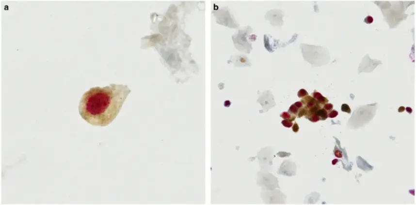
The p16/Ki-67 dual staining technique is a method that detects two important biomarkers. P16 and Ki-67. In a single tissue sample to provide detailed information, about cellular growth and possible cancerous changes. This special method uses both p16 and Ki-67 markers to understand cell proliferation p16 is a protein that stops certain enzymes related to cell division and is often increased in cells infected with HPV that can cause cancer; while Ki-67 is a protein found in cell nuclei linked to how fast cells are growing. By using these markers this way can help doctors identify abnormal cell growth more accurately.

How p16 and Ki-67 Markers Work Together
The synergy between p16 and Ki-67 markers lies in their ability to highlight distinct aspects of cellular activity. While p16 accumulation indicates potential viral oncogene activity, Ki-67 reflects the rate of cell division. By examining both markers simultaneously, clinicians can assess whether the observed cellular changes are indicative of high-risk lesions or benign conditions. This dual approach enhances diagnostic accuracy, enabling more informed clinical decisions.
The Diagnostic Value of p16/Ki-67 Dual Staining
The significance of using p16/Ki–67 staining as a diagnostic tool is most noticeable in screening for cervical cancer and other conditions linked to HPV infections. The technique helps in identifying abnormal cell changes and differentiating between temporary infections and those that have a higher likelihood of developing into cancerous conditions. Its usefulness is not limited to cervical issues but also holds promise in detecting abnormal tissue changes, in different parts of the body. For more detailed information on this technology, you can explore p16/Ki-67 Dual Staining.
Source from https://www.nature.com/articles/modpathol201716
Which Symptoms Are Indicative of p16 Positive Cases?
Common Symptoms Associated with p16 Positivity
Symptoms indicative of p16 positivity often correlate with the underlying pathology it represents. In cases of HPV-related lesions, common symptoms may include unusual bleeding or discharge, persistent pain, or visible changes in tissue morphology such as warts or lesions. These symptoms warrant further investigation through histological examination and dual staining techniques to confirm the presence of high-risk HPV activity.
How Symptoms Guide the Use of p16/Ki-67 Dual Staining
Clinicians rely on symptoms that appear consistently to determine when to use p16/Ki-67 dual staining as a diagnostic tool effectively in their practice. The identification of indicators like atypical squamous cells, in regular screenings prompts them to utilize this dual staining technique to evaluate both the extent and likelihood of progression associated with these observations. By integrating symptomatology with advanced staining techniques, healthcare providers can tailor their diagnostic approach to each patient’s unique presentation.
Solutions for Optimal Use
To maximize the benefits of p16/Ki-67 dual staining in clinical practice, it is essential to adopt optimized protocols that ensure accurate results. Leveraging solutions like those offered by Celnovte’s Solution Center can enhance the effectiveness of your diagnostic workflows. These solutions provide guidance on best practices for tissue preparation, staining procedures, and result interpretation—ensuring that you achieve reliable and reproducible outcomes every time.
By understanding how these biomarkers interact within pathological contexts and applying them judiciously based on patient symptoms, you can significantly enhance diagnostic precision and improve patient care outcomes.
How Can You Differentiate Between Benign and Malignant Conditions Using p16/Ki-67?
Identifying Benign Conditions with Dual Staining
p16/Ki-67 dual staining plays a pivotal role in distinguishing benign conditions from malignant ones. In benign cases, the expression of p16 may be present but typically without concurrent Ki-67 activity. This lack of proliferation marker Ki-67 indicates that while the cells might be under some stress or influence, they are not actively dividing at a high rate, suggesting a non-malignant nature. By examining these markers together, clinicians can confidently categorize lesions as benign when Ki-67 is absent or minimally expressed alongside p16.
Recognizing Malignant Patterns through Marker Analysis
In contrast, malignant conditions often exhibit a pattern where both p16 and Ki-67 are strongly expressed. This dual positivity suggests that the cells are not only under oncogenic stress but are also proliferating rapidly, a hallmark of malignancy. The simultaneous presence of these markers allows for precise differentiation between high-risk lesions and benign anomalies. Utilizing this dual staining technique enhances diagnostic accuracy by highlighting malignant transformation early in its course, thereby facilitating timely intervention.
Why Is Early Detection Crucial in Symptom Analysis with p16/Ki-67?
The Importance of Timely Diagnosis
Early detection through p16/Ki-67 dual staining is crucial because it enables healthcare providers to identify potentially malignant lesions before they progress to advanced stages. Timely diagnosis allows for more effective management strategies, reducing the likelihood of invasive disease development. By employing this method during initial symptom analysis, practitioners can intervene sooner, improving patient outcomes and potentially curbing the progression of precancerous conditions.
How Early Detection Impacts Treatment Outcomes
The impact of early detection on treatment outcomes cannot be overstated. When abnormalities are identified at an early stage using p16/Ki-67 dual staining, there is a broader range of therapeutic options available that can be less invasive and more effective. This proactive approach not only improves survival rates but also enhances the quality of life for patients by minimizing the need for aggressive treatments later on. For further insights into optimizing early detection methods, consider exploring Celnovte’s Solution Center.
By including p16/Ki-67 staining in regular diagnostic procedures we can thoroughly assess symptoms and distinguish between benign and malignant conditions more accurately This approach allows healthcare providers to customize patient care by combining this knowledge with each individual’s specific symptoms to meet their distinct clinical requirements accordingly.
Are There Challenges in Interpreting Results from p16/Ki-67 Dual Staining?
Potential Pitfalls in Result Interpretation
Analyzing findings from p16/Ki-67 dual staining poses difficulties that could influence diagnostic precision significantly. A key challenge involves the risk of positive or negative results due to technical challenges like mishandled tissues or inconsistencies in staining procedures. The variations in staining strength and marker presentation can further add complexity to understanding results. May result in misidentifying abnormalities. Recognizing these challenges is vital, for ensuring diagnoses and making informed clinical decisions effectively.
Strategies to Overcome Diagnostic Challenges
To tackle these obstacles effectively and ensure p16/Ki-67 dual staining assessments; it is crucial to implement various techniques for enhancing reliability measures. One vital approach involves standardizing protocols, for tissue preparation and staining procedures to reduce variations and enhance uniformity in results. Moreover; utilizing top-notch reagents and implementing quality control checks can help reduce technical errors significantly. Additionally, adopting advanced imaging techniques and software tools for precise quantification of marker expression can aid in distinguishing between benign and malignant patterns more accurately.
For those seeking comprehensive solutions to optimize their diagnostic workflows, Celnovte’s Solution Center offers valuable resources and guidance on best practices. By leveraging these solutions, healthcare providers can enhance their ability to interpret p16/Ki-67 dual staining results effectively, ultimately improving patient care outcomes.
Integrating these methods into practice not only tackles possible challenges but also guarantees that healthcare professionals have the essential resources to make well-informed decisions grounded in dependable data This forward-thinking method encourages a more precise evaluation of symptoms leading to prompt action and improved handling of HPV-associated conditions.
How Do Technological Advances Enhance the Accuracy of p16/Ki-67 Dual Staining?
Innovations in Staining Techniques
Technological advances have significantly refined staining techniques, enhancing the accuracy of p16/Ki-67 dual staining. One such innovation is the development of chromogenic in situ hybridization, which utilizes an HRP-DAB chromogenic system to detect RNA and observe staining results under a light microscope. This method is straightforward to implement and offers substantial clinical and pathological value, particularly in diagnosing viral infections like EBV. These advancements allow for more precise visualization of biomarker expressions, providing clearer insights into cellular activities.
Moreover, multiplex immunohistochemistry (mIHC) has emerged as a groundbreaking technique that enables the simultaneous detection of multiple biomarkers within a single tissue section. By optimizing staining and mounting strategies, researchers can achieve maximal signal preservation and superior image quality. This approach not only elevates diagnostic precision but also facilitates a deeper understanding of complex biological systems. For those interested in exploring advanced staining products, Celnovte’s offerings provide robust solutions for achieving high-quality imaging outcomes.
Leveraging Technology for Improved Diagnostic Precision
The integration of cutting-edge technology in p16/Ki-67 dual staining has revolutionized diagnostic precision. By leveraging advanced imaging systems and software tools, clinicians can accurately quantify marker expression levels and differentiate between benign and malignant conditions with greater confidence. High-performance multiplex immunofluorescence kits, such as those provided by CNT, deliver stronger signal intensities and clearer biomarker localization compared to traditional methods.
The advancements in technology guarantee outcomes and enduring fluorescence stability essential for trustworthy diagnoses, in healthcare settings. This enables medical professionals to make founded decisions using precise information that ultimately enhances patient care results. Incorporating these new technologies into daily procedures can elevate your diagnostic skills and help navigate the challenges of interpreting dual staining findings accurately.
In conclusion, technological advancements have played a pivotal role in enhancing the accuracy of p16/Ki-67 dual staining. Through innovative techniques and state-of-the-art tools, clinicians are better equipped to interpret complex biomarker interactions and provide tailored care based on precise diagnostic insights. For comprehensive solutions to optimize your diagnostic workflows, consider exploring Celnovte’s Solution Center, where you’ll find valuable resources designed to support your clinical practice.
Solutions for Effective Implementation of p16/Ki-67 Dual Staining
Solutions to Optimize Diagnostic Processes
Implementing p16/Ki-67 dual staining effectively requires a strategic approach to ensure that diagnostic processes are optimized for accuracy and reliability. By adopting solutions that streamline workflow and enhance result precision, healthcare providers can significantly improve their diagnostic capabilities. Utilizing well-defined protocols for tissue preparation and staining procedures is crucial in minimizing variability and achieving consistent outcomes.
Advanced imaging technologies and software tools play a pivotal role in optimizing these processes. They provide precise quantification of biomarker expression levels, aiding in the differentiation between benign and malignant conditions. By integrating these technological solutions into routine practice, clinicians can enhance their ability to interpret complex biomarker interactions accurately.
Ensuring Accurate and Reliable Results with p16/Ki-67 Dual Staining
For dependable outcomes using p16/Ki67 dual staining technique is crucial to emphasize quality control steps and utilize top-notch materials that deliver optimal performance results. Implementing quality assessments during staining procedures helps minimize technical mishaps that may result in incorrect positive or negative findings. Premium grade reagents guarantee signal strengths and distinct biomarker positioning essential for accurate diagnostic evaluations.
By leveraging cutting-edge multiplex immunofluorescence kits, such as those offered by CNT, clinicians can achieve superior signal-to-noise ratios and reproducible results. These kits are designed to provide long-term fluorescence stability, essential for reliable diagnosis over time.
For comprehensive guidance on implementing these best practices, healthcare providers can explore Celnovte’s Solution Center, which offers valuable resources tailored to support clinical workflows. By adopting these strategies, you can enhance diagnostic precision and improve patient care outcomes through the effective implementation of p16/Ki-67 dual staining techniques.
")