Understanding IHC: Key Uses in Modern Medical Diagnostics

By admin
The Basics of Immunohistochemistry (IHC)
Definition and Fundamentals of IHC
Immunohistochemistry (IHC) is a crucial analytical technique used in pathology laboratories worldwide. It involves the detection of antigens (proteins) in cells of a tissue section by exploiting the principle of antibodies binding specifically to antigens in biological tissues. This technique allows researchers and diagnosticians to visualize the distribution and localization of specific cellular components within tissue samples. By using colorimetric reactions or fluorescence, these bound antibodies can be detected and analyzed under a microscope, providing valuable information about the presence and quantity of specific proteins.
How IHC Works
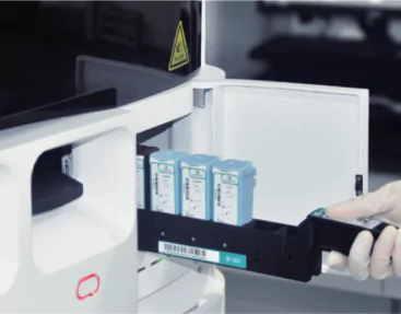
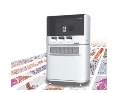
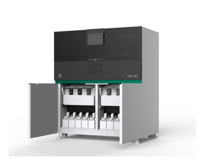
The process of IHC involves several steps to ensure precise and specific staining of the target antigens. Initially, tissue samples are collected and fixed to preserve cellular structures. These samples are then embedded in paraffin and cut into thin sections. The sections are mounted on glass slides and treated with a series of chemicals to unmask the antigens and facilitate antibody binding. Primary antibodies, which specifically bind to the target antigens, are applied, followed by secondary antibodies that are conjugated with enzymes or fluorescent dyes. These secondary antibodies amplify the signal, enabling the visualization of the antigen-antibody complex under a microscope. CNT330 Full Automatic Multiplex IHC Stainer, CNT300 Full Automatic Multiplex IHC Stainer, and CNT360 Full-automatic IHC&ISH Stainer are available machines.
Core Applications of IHC in Medical Diagnostics
Cancer Diagnosis
One of the primary applications of IHC is in the diagnosis and classification of cancer. IHC helps pathologists determine the tissue of origin of metastatic tumors and differentiate between benign and malignant lesions. By identifying tumor-specific markers, clinicians can classify the type of cancer and provide personalized treatment plans. For instance, the detection of Her2/neu protein is crucial in identifying breast cancer subtypes and guiding targeted therapy decisions. Furthermore, IHC is used to assess hormone receptor status in breast cancer, which is essential for determining the suitability of hormone therapy.
Infectious Disease Detection
Immunohistochemistry (IHC) is widely employed in detecting infectious diseases by identifying specific pathogens in tissue samples. This technique allows for the localization of viral, bacterial, and fungal antigens within infected tissues. For instance, IHC can identify human papillomavirus (HPV) in cervical cancer biopsies and tuberculosis bacteria in lung tissue samples. This aids in confirming infections’ presence, understanding the disease’s pathogenesis, and guiding suitable treatment strategies. The capability of IHC to precisely locate pathogens within tissues renders it a crucial tool in infectious disease diagnostics.
Neurological Disorders Identification
In the field of neurobiology, IHC plays a significant role in diagnosing and studying neurological disorders. By detecting specific neuronal and glial markers, researchers can uncover the underlying mechanisms of diseases such as Alzheimer’s, Parkinson’s, and multiple sclerosis. For instance, the identification of amyloid plaques and tau protein tangles through IHC is crucial in diagnosing Alzheimer’s disease. IHC also aids in the detection of neuroinflammatory responses and the presence of infectious agents in neurodegenerative diseases, providing insights into disease progression and potential therapeutic targets.
CNT330 Full Automatic Multiplex IHC Stainer
Celnovte is a leading biotech company that specializes in the development of advanced medical diagnostic tools. One of their flagship products is the CNT330 Full Automatic Multiplex IHC Stainer. This state-of-the-art machine is designed to automate the process of immunohistochemistry staining, a key technique used in modern medical diagnostics.
The CNT330 is capable of processing up to 60 slides in just 2.5 hours, making it an efficient solution for high-throughput laboratories. It supports the development of new technologies and projects such as chromogenic in situ hybridization, multi-color IHC, and p16/ki-67 staining.
The machine’s multi-color IHC capability enables the concurrent visualization of various antigen targets on a single slide. This significantly enhances the detection rate of tumor microinfiltration, thereby boosting the precision of pathological diagnosis.
Moreover, the CNT330 is compatible with a wide range of immunohistochemistry reagents. Celnovte offers more than 460 primary antibodies, independently developed secondary antibody detection systems, and over 200 quality control products.
The CNT330 also supports intraoperative rapid frozen IHC, which can complete staining operations within 15 minutes. This feature greatly improves diagnostic accuracy for atypical morphology patients, difficult cases, and young patients, reducing the rate of delayed diagnosis.
In conclusion, the CNT330 Full Automatic Multiplex IHC Stainer from Celnovte is a comprehensive solution for immunohistochemistry, offering high-quality staining results and supporting a wide range of diagnostic applications. It is an invaluable tool for any laboratory involved in modern medical diagnostics.
Advantages and Limitations of IHC
Strengths of IHC in Diagnostics
Immunohistochemistry (IHC) possesses several notable strengths that make it fundamental in modern diagnostics. One of its primary benefits is the capability to precisely localize antigens within tissue samples. This precise localization assists in the accurate diagnosis of various diseases, particularly cancers, by enabling detailed visualization of cellular-level changes. Another major strength of IHC is its versatility. The method can be utilized on a wide array of tissue types and is capable of detecting diverse antigens, ranging from proteins to small molecular compounds. Additionally, IHC can be performed using both fresh and archival formalin-fixed, paraffin-embedded tissues, enhancing its utility in clinical practices.
In addition, IHC is instrumental in prognostication and therapy guidance. By determining the expression of biomarkers, such as hormone receptors in breast cancer, IHC helps in predicting disease outcomes and tailoring personalized treatment plans. Furthermore, IHC has relatively high sensitivity and specificity when appropriate antibodies are used, providing reliable results that are crucial for effective patient management. Lastly, the visual nature of IHC allows pathologists to assess not just the presence of the target antigen but also the intensity and distribution pattern, offering deeper insights into disease mechanisms.
Challenges and Considerations
Despite its numerous advantages, IHC is not without limitations and challenges. One key consideration is the quality of antibodies used. Variability in antibody quality can lead to inconsistent staining results, affecting the accuracy of the diagnosis. This necessitates rigorous validation of antibodies and routine quality control procedures to ensure reliability. Another challenge is the potential for non-specific binding, which can result in background staining and complicate the interpretation of results. Optimizing the staining protocol and using appropriate controls can help mitigate this issue.
Additionally, IHC is often limited by the availability of specific antibodies for certain antigens, which can restrict its use in some diagnostic applications. The technique also requires specialized equipment and trained personnel, which may not be readily available in all laboratory settings, particularly in resource-limited regions. Furthermore, interpreting IHC results can be subjective, depending on the pathologist’s experience, which highlights the need for standardized scoring systems and automated image analysis tools to enhance consistency.
Innovations and Future Trends in IHC
Technological Advances Enhancing IHC
The field of immunohistochemistry (IHC) is constantly advancing, propelled by technological progress that boosts its capabilities and efficiency. A notable innovation in this area is the introduction of automated staining systems, such as the CNT330 Full Automatic Multiplex IHC Stainer. These systems optimize the staining procedure by reducing the need for manual intervention, thereby enhancing throughput and reproducibility. Additionally, automation reduces human error and ensures uniform application of reagents, resulting in more dependable outcomes.
Furthermore, multiplex IHC is an emerging technology that allows the simultaneous detection of multiple biomarkers on a single tissue section. This enables a more comprehensive analysis of the tissue environment and interactions between different cellular components. Emerging fluorophore conjugates and chromogenic substrates enhance the detection sensitivity and specificity, providing clearer and more detailed visualizations. Digital pathology and advanced imaging techniques, including artificial intelligence and machine learning algorithms, are being integrated with IHC to enable automated image analysis, quantification of staining intensity, and pattern recognition.
Emerging Applications in Personalized Medicine
IHC is poised to play a pivotal role in the advancement of personalized medicine. By characterizing the molecular profiles of tumors, IHC facilitates the identification of specific genetic and protein markers that can be targeted with individualized therapies. Personalized oncology relies on IHC to determine the expression of therapeutic targets such as PD-L1 in immunotherapy and Her2/neu in targeted breast cancer treatment. This precision approach maximizes therapeutic efficacy while minimizing side effects.
Moreover, IHC is being employed in the development of companion diagnostics, which are tests designed to accompany specific therapies to determine patient eligibility and monitor treatment responses. This application of IHC ensures that patients receive treatments that are most likely to be effective based on their unique biological markers. In addition to oncology, IHC is expanding its applications to other fields, including cardiology and infectious diseases, where it helps in identifying relevant disease markers and developing targeted interventions.
To sum up, as immunohistochemistry (IHC) technology progresses, it is poised to transform the realm of medical diagnostics by providing more advanced tools for disease detection and tailored treatments. With continuous innovations and an increasing comprehension of molecular biology, IHC will remain a key player in medical research and clinical practice, offering essential insights that lead to improved patient outcomes.


