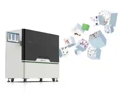
Slide Stainers in Cancer Research: Key Applications and Innovations
2025-02-13
By admin
Using slide stainers is crucial in cancer research as they automate tissue staining to ensure efficient diagnoses. This article explores how this technology enhances pathology labs and boosts the accuracy of cancer detection.
What Are Slide Stainers and How Do They Work in Cancer Research?
In the field of cancer research and diagnosis equipment dvancement is the emergence of slide stainers as an element in the process. The purpose of these machines is to automate the staining of tissues mounted on slides—a critical step in pathology exams. They use dyes or antibodies to emphasize various elements in cells or tissues to assist pathologists, in detecting potential cancer indicators.
The Role of Slide Stainers in Pathology Labs
In pathology labs, slide stainers are crucial as they simplify the staining process by automating it. This helps in minimizing errors made by humans and enhances efficiency by processing several samples. In cancer research where accuracy and time are key factors, slide stainers offer dependability and uniformity, guaranteeing precise and reproducible diagnostic outcomes.
Mechanisms of Slide Staining Technology
The science behind slide machines incorporates various processes that guarantee thorough and efficient staining results. It’s important to understand that these devices manage the application of stains using timing and temperature control methods. The precision in this process is crucial as it impacts how stains attach to cell parts and improves the clarity of structures when viewed through a microscope.
Importance of Precision and Accuracy in Cancer Diagnosis
Accurate and precise cancer diagnosis is essential for effective treatment outcomes. Slide stainers play a role in ensuring consistent staining results that aid in identifying cancer cells accurately. This precision is key in reducing diagnostic errors and enhancing patient care through early detection and treatment planning opportunities.
How Are Slide Stainers Revolutionizing Cancer Diagnosis Equipment?
The use of slide stainers in cancer diagnosis equipment has transformed the field by boosting capabilities with the help of technological advancements. These advancements have resulted in accuracy levels and faster processing times while also ensuring seamless integration with other technologies.
Enhancements in Diagnostic Accuracy
A notable progress is the enhancement in precision. Using automated systems allows for staining across samples, reducing potential variations that may cause misinterpretation. Such uniformity is crucial for ensuring cancer diagnoses.
Speed and Efficiency Improvements
Reduction in Turnaround Time
Using slide stainers can help speed up the processing of samples in pathology labs by cutting down on turnaround time through the automation of tasks. This enables faster diagnosis and treatment commencement.
Automation Benefits
The advantages of automation go beyond speed. They also involve less manual work and a decreased chance of human mistakes, resulting in improved efficiency in laboratory operations and more effective allocation of resources.
Integration with Digital Pathology
The merging of slide stainers with pathology platforms marks a significant advancement in technology usage in healthcare settings. This union enables the digitization and examination of stained slides digitally, enabling consultations and the application of sophisticated image analysis methods.
What Are the Key Innovations in Slide Staining Technology?
In the past few years, there have been significant advancements in slide-staining technology that aim to improve its use in cancer research.
Advances in Automated Slide Stainers
Features of Modern Automated Systems
Current automatic slide staining machines are designed with functionalities, like protocols and the ability to handle multiple reagents at once while also offering high throughput capabilities. This enables users to tailor the staining procedures to fit their research requirements.
Impact on Laboratory Workflow
The influence on how laboratories operate is significant since these systems boost productivity by minimizing the need for work and expanding the ability to process samples efficiently, enabling laboratories to manage higher sample volumes without sacrificing quality.
Development of Specialized Stains for Cancer Detection
Types of Specialized Stains
Researchers have created dyes to pinpoint specific indicators linked to different types of cancers effectively using specialized methods like p16/KI67 dual staining on cells or tissue samples, all in a single slide, for improved diagnosis of cervical precancerous lesions.
Application in Different Cancer Types
These specific dyes are used in types of cancer to detect distinct markers found in tumors with precision and accuracy. In particular, the p16/KI67 double stain method is commonly employed in screening for cancer due to its reliance on objective color interpretation results.
Microfluidic Slide Staining Systems
Cutting-edge advancements, in slide staining technology are demonstrated by systems that use small channels to accurately handle fluids. The systems provide control over stain application which results in more consistent staining and reduced use of chemicals.
How Does Celnovte Stand Out as a Reliable Slide Stainer Supplier?
When evaluating suppliers of slide-staining equipment for medical labs, Celnovte stands out as a standout choice thanks to its dedication to excellence and cutting-edge solutions in the field of cancer diagnosis technology. It’s worth delving into the ways Celnovte sets itself apart in the market of providers offering equipment for diagnosing cancer.
Quality Assurance and Reliability
At Celnovte we prioritize top-notch quality in our slide stainers to ensure accurate diagnostic results are maintained effectively. Their products go through testing to align with industry standards, giving you dependable tools that improve the precision of laboratory work.
Technological Innovations
With the technology embedded in their slide stainers, Celnovte stays ahead in cancer research progressions. Their products meet laboratory requirements by including programmable protocols and the ability to use multiple reagents.
Customer Support and Training
Celnovtes customer service stands out for its support system and training initiatives that play a key role in ensuring laboratory personnel are well equipped to make the most of the equipment for efficient cancer research purposes.
Customization Options
At Celnovte offer customization features tailored to meet specific research needs. This adaptability enables you to adjust the slide staining procedure, for cancer research projects hence improving the significance and accuracy of your diagnostic results.
Conclusion
In cancer research, today slide stainers play a role in transforming how biological tissues are examined by scientists and medical professionals around, utilizing these devices to analyze specimens efficiently in pathology laboratories, leading to quicker results with higher precision levels with advancements such as automated procedures and custom stains like dual p16/KI67 staining, which identifies two biomarkers in cells or tissue samples from just a single slide, thereby helping significantly in identifying precancerous lesions in cervical areas. Their utility gets further elevated when combined with digital pathology technology, allowing for more sophisticated imaging methods to be employed, enhancing their overall effectiveness and usage within this field of study.
Celnovte distinguishes itself as a supplier through its products supported by cutting-edge technology and top-notch customer service. Their dedication, to ensuring quality control guarantees that labs using their slide stainers can perform cancer research with enhanced accuracy confidently.
FAQs About Slide Stainers in Cancer Research
Q1: What is the significance of p16/Ki-67 double staining in cancer diagnosis?
The p16/Ki67 double staining method identifies both p16 and Ki67 biomarkers in cells. Tissue samples, on a single slide to aid in diagnosing cervical precancerous lesions efficiently. It is commonly employed in cervical cancer screening because it relies on color interpretation outcomes rather than being influenced by subjective factors.
Q2: How do automated slide stainers improve laboratory workflow?
Automated slide-staining machines improve the efficiency of laboratories by reducing the need for tasks and increasing the capacity to process samples while minimizing mistakes made by humans. This results in operations and faster delivery of diagnostic findings.
Q3: Why is integration with digital pathology important for slide stainers?
Integration with pathology enables the utilization of digital imaging and examination of stained slides, for remote consultations and advanced image analysis methods—an advancement that boosts diagnostic capabilities and fosters collaborative research endeavors.


