
-
-
SHOP

Practical Overview of Multiplex Immunohistochemistry using TSA
![]() 2024-11-26
2024-11-26
By admin
In the realm of studies, it’s crucial to grasp the intricate dance between cells and proteins in tissues. Picture having the ability to see biomarkers at once within a single tissue sample revealing a treasure trove of insights beyond what traditional staining techniques can provide. That’s where the magic of multiplex immunohistochemistry (mIHC) shines. This innovative method enables the tagging of multiple biomarkers, on one tissue section simplifying a thorough examination of cell characteristics and their connections. Advances in mIH technology are revolutionizing areas such as cancer studies and immunology by offering researchers an understanding of intricate biological networks and structures. It serves as a resource for exploring the immune environment of tumors and uncovering the spatial connections among different cell categories, in specific sites.
The Power of TSA in mIHC
Tyramide signal amplification (TSA) is a pivotal technology that makes mIHC a reality. This new method improves the identification of targets, in samples effectively showing different biomarkers on a single tissue slice.TSA can smoothly blend with immunohistochemistry (ICH) offering more detailed protein data and boosting assay accuracy and sensitivity.
How TSA Works
It’s like a detective searching for fingerprints to uncover hidden clues. That’s similar to what TSA does but, on a tiny scale instead of traditional chromogenic IHC using horseradish peroxidase (HRP) to create a colored precipitate; TSA-based IHC generates a brighter fluorescent signal that is much more sensitive.
1. Antibody Binding: It starts with a primary antibody that specifically targets the protein of interest. Next, an HRP-conjugated secondary antibody binds to this primary antibody, forming a complex.
2. Tyramide Activation: The HRP enzyme acts like a catalyst, converting a fluorescently labeled tyramide substrate into a highly reactive form. This reactive tyramide swiftly attaches to tyrosine residues on nearby proteins, ensuring precise and localized labeling.
3. Covalent Bonding: Think of this step as locking the evidence in place. The reactive tyramide forms strong covalent bonds with tyrosine residues near the target protein. This permanent bond ensures that the fluorescent label remains attached even after the antibodies are removed.
4 . Antibody Removal: The antibodies are gently washed away, leaving behind only the brightly glowing fluorescent label attached to the target protein.
5. Multiplexing: Now, imagine repeating this process with different primary antibodies, each targeting a different protein, and using different colored fluorescent labels. This is the essence of multiplexing – the ability to visualize multiple targets on a single tissue section.
Figure 1: Working principle of TSA-based multiplex immunofluorescence
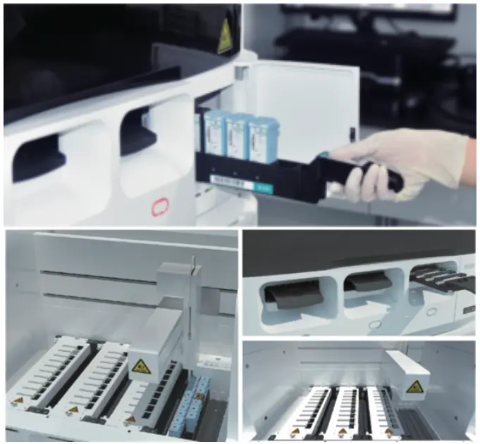
Celnovte Biotech is a leading provider of mIHC solutions, offering a comprehensive suite of products and services to empower your research. Explore their innovative solutions, including the CNT330 Full Automatic Multiplex IHC Stainer, designed to streamline your workflow and deliver exceptional results.
Applications of mIHC in Cancer Research and Biomarker Identification
The importance of multiplex data in cancer research and biomarker identification continues to grow. mIHC is proving to be an invaluable tool in these fields:
Understanding the Tumor Microenvironment: Examining a tumor is similar to observing an ecosystem in action through mIHC technology which enables scientists to observe the detailed connections between tumor cells and nearby immune cells revealing valuable information about tumor growth and response, to treatment protocols.
Patient Stratification for Immunotherapy: Molecular imaging in healthcare can assist in pinpointing individuals who could potentially see outcomes from particular immunotherapy treatments by examining the arrangement of immune indicators, within the tumor’s surrounding environment.
Biomarker Discovery: Imagine trying to find a needle in a vast haystack! mIHc makes it possible for scientists to study potential biomarkers all, at once in a single tissue sample – hastening the identification of fresh diagnostic and predictive indicators.
Development of New Cancer Therapies: A deeper understanding of the tumor microenvironment, facilitated by mIHC, can guide the development of more effective and targeted cancer therapies.
Advantages of TSA-Based mIHC
TSA-based mIHC surpasses traditional IHC methods in several key aspects:
Increased Sensitivity: Enhancing the signal is akin to increasing the volume of a transmission signal. TSA significantly boosts the signal strength to enable researchers to identify proteins, with low levels of abundance that could easily be overlooked otherwise.
Multiplexing Capability: Picture having the ability to hear musical instruments playing in an orchestra all at once! Through TSA-based mIlHC techniques, researchers can now label targets on a single tissue slice to conduct a more thorough and detailed analysis.
No Species Restriction for Primary Antibodies: Since the antibodies are taken out after each staining cycle is completed you can opt for antibodies, from the same species which simplifies the process of selecting antibodies and designing experiments.
Overcoming Challenges in TSA-Based mIHC Assays
While TSA-based mIHC is a powerful tool, researchers often encounter challenges:
Manual Steps: Traditional TSA protocols can be tedious, involving numerous manual steps that increase the risk of human error and variability.
Long Protocol Times: The multiple rounds of staining and washing can stretch out protocols, delaying results and impacting research efficiency.
High Reagent Consumption: TSA reagents can be expensive, and the multiple rounds of staining required can lead to significant reagent consumption, especially during the optimization phase.
Optimization Complexity: Perfecting TSA-based tests entails striking the balance of conditions for every target—a task that may be time-intensive and necessitate several rounds of fine-tuning.
Automation: The Solution for Streamlining TSA Workflows
Imagine having a robotic assistant to handle the repetitive tasks in your lab, freeing you up to focus on the bigger picture. That’s the power of automation in mIHC. Automated platforms, such as those offered by Celnovte Biotech, address the challenges of manual TSA protocols, reducing hands-on time, enhancing data quality and reproducibility, and minimizing reagent consumption.
Celnovte’s Solution provides detailed information on their automated staining platforms and how they can streamline your mIHC workflow.
Benefits of Automation:
High Signal-to-Background Ratio: Automation ensures precise reagent delivery and thorough washing steps, resulting in clearer signals with minimal background noise.
Uniform Staining: Automated platforms provide consistent staining across the entire tissue section, eliminating gradients and ensuring even signal distribution.
Efficient Antibody Elution: Effective removal of antibodies between staining cycles is crucial to prevent cross-reactivity and ensure accurate results. Automated systems achieve over 99% elution efficiency, guaranteeing reliable data.
Preservation of Tissue Morphology: Gentle automated handling minimizes tissue damage, preserving the morphology of the sample for reliable analysis.
Reproducibility: Automation eliminates human variability, leading to consistent and reproducible results across multiple experiments.
Reduced Turnaround Time: Automating time-consuming tasks, such as staining and washing, allows researchers to obtain results faster.
Reduced Reagent Consumption: Automated systems utilize precise dispensing mechanisms, minimizing reagent waste and reducing costs, especially during the optimization phase.
For example, the LabSat automated tissue stainer, highlighted in the sources, enables automation of IHC and IF assays using readily available reagents for reproducible and high-quality data. A 6-plex TSA staining can be completed in just four and a half hours using this platform.
Conclusion
Hospitals using TSA technology are introducing a cutting-edge method called mIHc that is transforming the field of research by enabling the visualization of multiple biomarkers simultaneously in one tissue sample. This innovative approach provides new perspectives on intricate biological processes. Although the standard mIHc protocols based on TSA can be tricky to implement the use of automation presents ways to simplify processes optimize data accuracy and lower expenses. Companies like Celnovte Biotech are driving this revolution, providing cutting-edge mIHC solutions to empower researchers in their quest to understand disease and develop new therapies.
FAQ
Q: What are the key advantages of using mIHC over traditional IHC methods?
A: mIHC offers several advantages over traditional IHC methods, including:
Increased sensitivity, allowing for the detection of low-abundance proteins.
Multiplexing capability, enabling the simultaneous analysis of multiple targets on a single tissue section.
No species restriction for primary antibodies, simplifying antibody selection and experimental design.
Q: What are the main challenges associated with TSA-based mIHC assays, and how can they be addressed?
A: The main challenges include:
The high number of manual steps involved makes the process time-consuming and prone to human error.
Long protocol times due to the multiple rounds of staining and washing steps.
High reagent consumption, which can be costly, especially during the optimization phase.
Complexity of optimization, requiring multiple cycles to find the optimal staining conditions.
Automation is the key to addressing these challenges, offering benefits such as:
Reduced hands-on time
Improved data quality and reproducibility
Minimized reagent consumption
Simplified optimization
Q: How is mIHC being used in cancer research and biomarker identification?
A: mIHC is being used to:
Understand the complex interactions between tumor cells and the surrounding immune cells within the tumor microenvironment.
Identify patients who are most likely to respond to specific immunotherapies based on the spatial distribution of immune markers.
Accelerate the discovery of new biomarkers by analyzing multiple potential targets simultaneously.
Guide the development of more effective and targeted cancer therapies based on a deeper understanding of the tumor microenvironment.
Q: What does the future hold for mIHC?
A: The outlook for mIHC looks bright as automation continues to progress and enhance its abilities in the future. With the expectation of creative uses being developed in the field of mIHC, it is likely to strengthen its contribution, to advancing our knowledge of diseases and enhancing patient treatment.