
-
-
SHOP

Periodic Acid-Schiff (PAS) Staining: A Histopathology Essential for Detecting Carbohydrates and Beyond
![]() 2024-11-21
2024-11-21
By admin
In the field of histopathology. The study of tissues under a microscope. Having visualizations is crucial for analysis purposes. Staining methods play a role in emphasizing particular elements within cells and tissues by making them visible when viewed through a microscope. A notable technique in this realm is the Periodic Acid-Schiff (PAS) stain, known for its effectiveness, in highlighting carbohydrates in tissues and uncovering structures that contribute to understanding various diseases better.
The Chemistry of PAS Staining: A Dance of Oxidation and Color
The PAS staining method uses a sequence of reactions that target carbohydrates in a way that makes them visible as brightly stained structures in the end. It all starts by applying periodic acid (HIO4) a strong oxidizing agent to tissue sections. This acid focuses on nearby hydroxyl groups (-OH) within carbohydrates like those, in 1,2-glycol groups, breaks their carbon-carbon bond, and creates two aldehyde groups (-CHO).
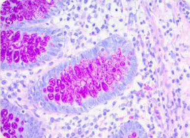
Schiff’s reagent, a colorless solution containing basic fuchsin and sulfur dioxide, then enters the stage. When Schiff’s reagent encounters the aldehyde groups generated by periodic acid oxidation, it undergoes a dramatic transformation. The sulfur dioxide component is released, and the basic fuchsin, now in its leuco form, binds to the aldehyde groups. This binding results in the formation of a vibrant magenta-colored compound, effectively painting the carbohydrate-rich structures within the tissue.
The level of magenta color intensity corresponds to the amount of carbohydrates in a semi-measurable way. Areas that contain high levels of carbohydrates will display a deep magenta stain while those, with lower carbohydrate levels will exhibit a lighter tint.
Performing PAS Staining: A Step-by-Step Guide
1. Deparaffinization and Hydration
- Immerse tissue sections in xylene to remove paraffin.
- Gradually rehydrate sections by passing them through decreasing concentrations of alcohol (100%, 90%, 80%, 70%) and water.
2. Oxidation
- Treat sections with a periodic acid solution (typically 1%) for 5-10 minutes.
3. Rinsing
- Rinse sections thoroughly with distilled water to remove residual periodic acid.
4. Schiff’s Reagent Treatment
- Immerse sections in Schiff’s reagent for 10-15 minutes.
5. Rinsing
- Wash sections under running tap water for 5-10 minutes to intensify the magenta color and remove unbound Schiff’s reagent.
6. Counterstaining
- Stain sections with hematoxylin for 15 seconds to highlight nuclei.
- Differentiate and blue the hematoxylin stain.
7. Dehydration, Clearing, and Mounting
- Dehydrate sections through increasing concentrations of alcohol.
- Clear sections with xylene.
- Mount sections on glass slides using a mounting medium like DPX.
Tips for Optimal PAS Staining
- Use commercially prepared periodic acid and Schiff’s reagent for quality and convenience.
- Store Schiff’s reagent in a dark, cool environment, protected from UV light and heat.
- Observe the color of the rinse water after Schiff’s reagent treatment; continue rinsing until the water runs clear.
Applications of PAS Staining: Illuminating a Spectrum of Diseases
The versatility of PAS staining lies in its ability to highlight a variety of carbohydrate-rich structures, making it a valuable tool in diagnosing and understanding a wide range of conditions.
1. Glycogen Storage Diseases: Revealing Metabolic Imbalances
The body’s main storage form of glucose is glycogen—a carbohydrate structure that can be affected by genetic metabolic conditions called glycogen storage diseases which disrupt its usual processing and cause abnormal buildup in different tissues of the body significantly impacting health conditions and functions, liver especially highlighted through PAS staining for diagnosis purposes.
2. Basement Membranes: Assessing Integrity in Renal Disease
Basement membranes are layers of extracellular matrix that offer structural support and function as selective barriers for epithelial and endothelial tissues. The PAS staining method is commonly employed to observe basement membranes visually and evaluate their thickness and condition. This technique proves valuable in the diagnosis of conditions since changes, in the thickness of the glomerular basement membrane may signify an underlying medical issue.
3. Fungal Infections: Identifying Carbohydrate-Rich Invaders
Many pathogenic fungi, such as Cryptococcus neoformans, possess cell walls rich in carbohydrates. These carbohydrate-rich walls become their downfall when confronted with PAS staining. The stain vividly highlights the fungal cell walls, aiding in their identification and differentiation from surrounding tissues.
4. Mucin: Characterizing Secretions in Health and Disease
Mucin is a type of glycoprotein that’s crucial for creating the protective mucus layer present in various body cavities. The PAS stain is utilized to help identify and describe mucin in tissues for staining purposes and provides insights, into the production of mucus by cells. This staining method is effective in distinguishing between acidic forms of mucins, which can help diagnose conditions linked to changes in mucin production.
5. Autoimmune Skin Diseases: A Potential Alternative to DIF
According to studies, PAS staining may offer a more cost-effective option, than DIF for diagnosing autoimmune skin conditions by illuminating carbohydrate-rich immune complexes. Studies have shown a strong correlation between PAS and DIF staining patterns in several autoimmune skin diseases, suggesting its potential as a valuable diagnostic tool.
Celnovte: A Trusted Name in Immunohistochemistry
Celnovte Biotech founded in 2010 is a supplier of top-notch immunohistochemistry (IHC) products focused on enhancing precise diagnostics in the oncology realm. Their dedication to excellence is evident in their array of IHC reagents and equipment such, as automated staining systems diverse primary antibodies, and specialized IHC kits that cater to the needs of the field.
The proficiency of Celnovte in IHC shines through its creation of staining technologies such as the MicroStacker™ Polymer Detection System that improves sensitivity and accuracy for better staining outcomes. Their dedication, to advancing research and development is showcased by their primary antibody development platform which has generated specialized antibodies targeting various subjects extensively.
Celnovte’s products have obtained ISO9001, ISO13485, and EU CE-ID certifications, demonstrating their adherence to international quality standards. Their commitment to providing comprehensive solutions for tumor pathology diagnosis has made them a trusted partner for researchers and clinicians worldwide.
Conclusion
The striking magenta shades of Periodic Acid Schiff staining have made it a crucial technique in the field of histopathology. This method not only reveals the complexities of glycogen storage disorders but also assists in identifying fungal infections. PAS staining remains a tool for exploring the unseen details of various health conditions, on a microscopic level.
 Staining Kit")
