Optimizing mIHC Techniques: Strategies for High Precision Results
2024-09-03
By admin
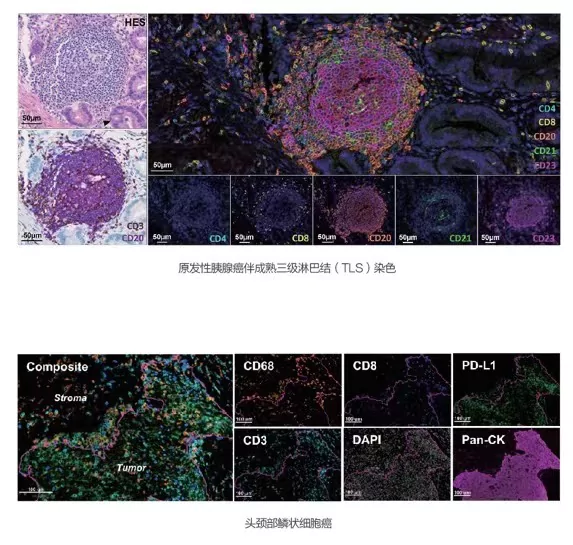
Multiplex immunohistochemistry (mIHC) plays a crucial role in enhancing our understanding of complex biological systems, particularly oncology. The ability to visualize multiple biomarkers simultaneously allows for the advanced classification of cancer types and subtypes, providing insight into tumor microenvironments and disease progression. By refining staining protocols and employing innovative strategies, researchers can maximize the potential of mIHC in both clinical and research settings.
Importance of Multiplex Immunohistochemistry (mIHC)
The importance of mIHC lies in its capacity to reveal the spatial and temporal dynamics of protein expression in tissue samples. This technology offers a detailed look at the cellular composition of tumors, aiding in the identification of therapeutic targets and prognostic markers. Furthermore, mIHC facilitates a better understanding of the interactions between different cell types within the tumor microenvironment, which is essential for developing effective cancer therapies. The simultaneous detection of multiple targets contributes to a comprehensive analysis, enabling a more integrated approach to cancer diagnosis and treatment planning.
Enhancing Cancer Classification through TSA-based mIHC Assays
One of the forefront techniques in mIHC is the use of tyramide signal amplification (TSA)-based assays, which significantly improve signal intensity and detection sensitivity. This methodology enhances the visualization of low-abundance proteins, often overlooked in traditional immunohistochemical approaches. By incorporating TSA in the staining protocol, researchers can achieve accurate identification and classification of cancer cells, leading to improved patient stratification and personalized treatment strategies. The robust performance of TSA-based assays in mIHC also allows for quantifiable data generation, which is essential for clinical validation and standardization in cancer diagnostics.
Best Practices for Multiplex Immunohistochemical Staining
To achieve high-quality results in mIHC, adherence to best practices in multiplex immunohistochemical staining is essential. Standardization of the staining protocol minimizes variability, ensuring reproducibility across experiments. Key considerations include the choice of primary antibodies, buffer systems, and incubation times, all of which should be optimized for the specific targets of interest. Implementing a systematic approach, such as using a Multiplex Immunohistochemical (mIHC) Kit designed for specific applications, can streamline the process, minimize user errors, and enhance outcomes.
Standardized Quantitative Analysis in mIHC/IF
Standardized quantitative analysis is a critical aspect of mIHC that allows for consistent interpretation of results. Utilizing automated image analysis systems can facilitate the objective quantification of staining intensity and cellular distribution of targets. By incorporating rigorous controls and calibrating the imaging equipment, researchers can improve the reliability of their quantitative assessments. This is particularly important when comparing results across different studies or patient samples, as variability in analysis can obscure meaningful biological differences. Through robust quantitative analysis, the clinical relevance of mIHC findings can be effectively communicated.
Application of Image Analysis in mIHC
Image analysis techniques are integral to the success of mIHC as they enhance the ability to interpret complex datasets derived from histological samples. The implementation of advanced software tools allows for the automated processing of images, facilitating the detection and quantification of multifaceted patterns of protein expression. These tools can differentiate signal from noise, enabling accurate identification of positive staining within heterogeneous tissues. Enhancing image analysis capabilities optimizes the data obtained from mIHC, providing deeper insights into cellular behavior and interactions.
Use of Landmark Markers for Tissue Indication
The use of landmark markers for tissue indication is an effective strategy in mIHC that aids in the localization of proteins of interest within the tissue architecture. By employing specific markers that delineate various tissue compartments, researchers can anchor their analyses, ensuring that protein expression is contextually relevant. This approach not only enriches the data obtained from mIHC but also enhances the biological interpretation of results. Landmark markers provide a spatial framework, making it easier to relate immunohistochemical findings to underlying pathology and clinical outcomes.
In conclusion, optimizing mIHC techniques requires a multifaceted approach that embraces technological advancements while adhering to established best practices. By leveraging TSA-based assays, ensuring standardized protocols, and applying sophisticated image analysis techniques, researchers can obtain high-precision results that significantly contribute to the advancement of cancer diagnostics and therapeutics. Continued innovations in mIHC will undoubtedly pave the way for enhanced understanding and treatment of various diseases, underscoring its critical role in contemporary biomedical research.
Technologies and Systems for Improved mIHC Performance
Celnovte
Celnovte stands as a premier biotechnology company, pioneering the development and production of sophisticated medical diagnostic tools. Driven by a relentless commitment to innovation, the company continually forges ahead, crafting groundbreaking solutions that meet the ever-evolving demands of contemporary medical diagnostics.
Celnovte represents a noteworthy advancement in mIHC technology by introducing innovative solutions for multiplex staining and visualizing protein interactions in tissues. Their cutting-edge platform enhances the sensitivity and specificity of mIHC, making it easier to identify multiple biomarkers within a single sample. The integration of proprietary reagents and optimized protocols can lead to improved signal intensity and reduced background noise, ultimately resulting in clearer and more interpretable images. Celnovte’s commitment to refining mIHC techniques contributes significantly to the broader adoption of complex multiplexing strategies in both research and clinical diagnostics.
Multiplex Immunohistochemical (mIHC) Kit
Advantages
With the ability to detect 6-8 markers simultaneously, the multiplex immunohistochemical (mIHC) kit eliminates species restrictions on antibody selection, allowing researchers to explore a broader range of biomarkers. This feature enables researchers to obtain more biological information from precious samples, enhancing the value of their experiments. Additionally, the kit improves target detection sensitivity and obtains specific image signals, ensuring accurate and reliable results. Furthermore, it achieves automated detection with high efficiency, good stability, and repeatability, streamlining the immunohistochemical process and reducing the potential for human error. Overall, Celnovte’s Multiplex Immunohistochemical Kit is a powerful tool that empowers researchers to gain comprehensive insights into complex cellular interactions and tissue architecture.
Applications
Celnovte’s Multiplex Immunohistochemical (mIHC) Kit has widespread applications across various fields of medical research and development. In drug development, the kit plays a crucial role. For preclinical studies, it enables functional evaluation of drugs by providing pathological analysis of the mechanism of action (MOA) substance. This critical information informs the development process and guides optimization. As clinical trials progress, the mIHC Kit assists in diagnosis by optimizing patient inclusion criteria, ensuring that trials are targeted and effective. Furthermore, it aids in developing accompanying diagnostic reagents, crucial for monitoring the efficacy and safety of drug candidates.
In clinical research, the Celnovte mIHC Kit contributes significantly to tumor characterization and understanding. It facilitates molecular classification of tumors, aiding in tumor immune-assisted disruption strategies. Additionally, the kit is invaluable in exploring tumor heterogeneity, providing insights into prognosis and risk assessment of metastasis. This knowledge is crucial for personalizing treatments and improving patient outcomes.
Finally, the Celnovte mIHC Kit serves as a powerful tool in basic scientific research. It enables in-depth investigation into the mechanism of immune checkpoints, revealing the expression, typing, and localization of immune control proteins. This fundamental understanding informs the development of new therapies and enhances our ability to manipulate the immune system. Furthermore, the kit allows for detailed analysis of the tumor microenvironment, shedding light on the interplay between cancer cells and their surroundings. By providing in situ analysis of various biomarkers within tissues, researchers can gain invaluable insights into tumor biology and progress towards new treatments.
Future Directions and Potential Developments in mIHC Protocols
Emerging Trends in Spatial Biology and Their Impact
Spatial biology is an evolving field that holds the promise of further enhancing mIHC techniques by integrating advanced imaging methods and spatially-resolved transcriptomics. These trends enable researchers to obtain a more detailed understanding of cellular interactions in their native contexts and explore patterns of protein expression within tissue architecture. As spatial biology develops, it will likely support the identification of new biomarkers that reflect the complexity of tissue microenvironments, thereby driving the next generation of mIHC applications. Furthermore, the fusion of mIHC with other modalities like single-cell sequencing may offer insights into tumor heterogeneity and facilitate the discovery of novel therapeutic approaches.
Continuous Refinement of Multiplex Immunohistochemical (mIHC) Kits
The continuous refinement of Multiplex Immunohistochemical (mIHC) Kits is essential for improving their performance and expanding their applications. Ongoing research focuses on optimizing antibody combinations to maximize specificity and sensitivity, reducing issues related to cross-reactivity. Additionally, improvements in reagent technology, including the development of new detection systems and signal amplification strategies, are crucial for enhancing the overall robustness of mIHC protocols. Regular updates to these kits will ensure that researchers have access to the latest advancements and can confidently utilize mIHC for high-quality, reproducible results in their studies. As such, manufacturers play a significant role in fostering innovation and facilitating the widespread adoption of optimized mIHC methodologies within the scientific community.
In summary, advancing the capabilities of mIHC through the use of technologies like Celnovte and refining Multiplex Immunohistochemical (mIHC) Kits will contribute to the accuracy and comprehensiveness of results obtained from multiplex staining. By embracing emerging trends in spatial biology and continuously improving methodologies, researchers can leverage mIHC’s full potential to enhance cancer diagnostics and yield deeper insights into complex biological systems. Collectively, these innovations form the basis for future breakthroughs in the understanding and treatment of various diseases, underscoring the critical importance of optimizing mIHC techniques in contemporary biomedical research.


