
-
-
SHOP

Navigating the Complexities of Multiplex Immunohistochemistry (mIHC): A Comprehensive Guide to Key Influencing Factors
![]() 2024-12-03
2024-12-03
By admin
Multiplex immunohistochemistry (mMIHC) has become a method in the field of biomedical research by allowing researchers to observe and study various biomarkers within a single tissue sample simultaneously. This advancement provides insights into the spatial connections and interactions among cells in a tissue section leading to a more profound comprehension of intricate biological processes especially in the realms of cancer research and immunology. However achieving consistent results, with mMIHC requires thorough attention to numerous factors at every stage of the experimental process from preparing samples to interpreting data.
Sample Preparation: Laying the Groundwork for Success
As with any scientific endeavor, the foundation of reliable mIHC results lies in meticulous sample preparation. The sources emphasize the importance of high-quality tissue sections, highlighting that thin, flat sections properly adhered to charged slides are essential for obtaining uniform and consistent staining, minimizing variability, and ensuring the integrity of subsequent analyses.
Tissue Fixation: Preserving the Blueprint of Life
Preserving samples in a lifelike state through tissue fixation is an important initial stage in the mIHC workflow process. The selection of type and duration of fixation along with temperature conditions can greatly influence the maintenance of tissue structure and the accessibility of target epitopes for antibody attachment. Formalin is a utilized fixative that has the potential to create protein cross-links which may obscure epitopes and impede antibody binding. As a result, it is essential, to fine-tune fixation procedures. Maintain consistency across samples to reduce variability and ensure accurate staining outcomes.
Section Adhesion: Securing the Canvas for Staining
Proper adhesion of tissue sections to slides is paramount for preventing detachment during staining procedures, which can lead to uneven staining, loss of tissue, and ultimately, compromised results. While protein-based adhesives might be tempting for securing sections, they can interfere with antibody binding, leading to inconsistent staining and difficulties in interpretation.
Antigen Retrieval: Unveiling the Hidden Treasures
The process of antigen retrieval plays a role in both IHC and mIHC techniques when working with formalin-fixed tissues. The use of formalin for tissue preservation can sometimes hide target epitopes due to protein cross-linking caused by the fixation process. This interference can lead to challenges in antibody binding and produce faint or non-staining on the tissues. Antigen retrieval techniques are designed to counteract this masking effect by revealing the epitopes and making them accessible, for antibody binding. This process ultimately enhances the intensity and accuracy of staining results.
Heat-Induced Epitope Retrieval (HIER): A Thermal Dance to Unlock Epitopes
HIER, the most commonly employed antigen retrieval technique, involves subjecting tissue sections to elevated temperatures in specific retrieval buffers. This process utilizes heat to break the cross-links formed during fixation, exposing the hidden epitopes. HIER can be performed using various heating methods, including steamers, microwaves, pressure cookers, or dedicated HIER instruments. Optimizing the buffer and heating settings is crucial for each antibody and antigen combination to achieve retrieval while preserving tissue integrity ensuring optimal results without damage to the sample structure and composition during HIER process commonly using buffers like citrate (at pH 6) EDTA (at pH 8) and Tris-EDTA (at pH 9).
Protease-Induced Epitope Retrieval (PIER): Enzymatic Precision for Epitope Exposure
PIER is a method for antigen retrieval that involves using proteolytic enzymes such as proteinase K, pepsin, and trypsin to break down cross-linked proteins and reveal hidden epitopes within tissues. However, PIER needs to be fine-tuned carefully since much enzymatic treatment can cause damage to tissues and disrupt their structure leading to changes, in staining quality and interpretation.
Blocking: Silencing the Noise for Crystal Clear Signals
Blocking is a part of mIHC to prevent antibodies, from sticking to the wrong parts of tissues instead of the target areas; this can cause extra background noise and make it harder to interpret the data accurately by creating false positives in the results.
Endogenous Enzyme Blocking: Taming the Unwanted Catalysts
Some tissues naturally contain enzymes that can interfere with the detection system used in mIHC, particularly those relying on enzymatic reporters like horseradish peroxidase (HRP). These endogenous enzymes can lead to false-positive signals and elevated background noise. Therefore, blocking these endogenous enzymes is essential for ensuring accurate and specific staining. For instance, in chromogenic detection systems employing HRP, endogenous peroxidase activity can be blocked using hydrogen peroxide or other specific inhibitors.
Autofluorescence: Quelling the Inner Glow
Some types of tissue components naturally emit light when exposed to specific wavelengths due to autofluorescence characteristics. This can create background interference and make it difficult to detect specific fluorescent signals. To address this issue, specialized blocking reagents or quenching techniques are used. These reagents either attach to autofluorescent molecules to hide their fluorescence or use chemical reactions to minimize autofluorescence. Choosing the right blocking reagents and optimizing them is vital for obtaining the best signal-to-noise ratios, in fluorescent mIHC experiments
Non-Specific Antibody Binding: Guiding Antibodies to their True Destination
Reagents known as blocking agents are used to prevent antibodies from binding to proteins or tissue elements by utilizing a mix of proteins like bovine serum albumin (BSA) or regular serum that attach to possible non-specific binding sites and prevent antibodies, from adhering to them so they can mainly target their intended epitopes.
Antibody Selection and Validation: The Cornerstones of Specificity
At the heart of successful mIHC lies the selection and rigorous validation of high-quality antibodies. The specificity and sensitivity of the chosen antibodies will directly impact the staining results, influencing data quality and the ability to draw meaningful conclusions.
Primary Antibodies: The Spearhead of Target Recognition
Primary antibodies are the first line of attack in mIHC, specifically targeting the biomarkers of interest within the tissue section. The choice between monoclonal and polyclonal antibodies is a critical decision, each offering distinct advantages and potential drawbacks.
Celnovte: Expanding the Palette with Multiplex Fluorescence IHC Kits
Celnovte offers a comprehensive range of multiplex fluorescence IHC kits and single-color TSA dyes, providing researchers with a versatile toolkit for fluorescent mIHC experiments. These kits can be seamlessly integrated with their innovative MicroStacker™ Polymer Detection System, a next-generation solution for IHC detection featuring enzyme-labeled antibodies and advanced polymer technology for enhanced sensitivity and specificity in both manual and automated IHC applications.
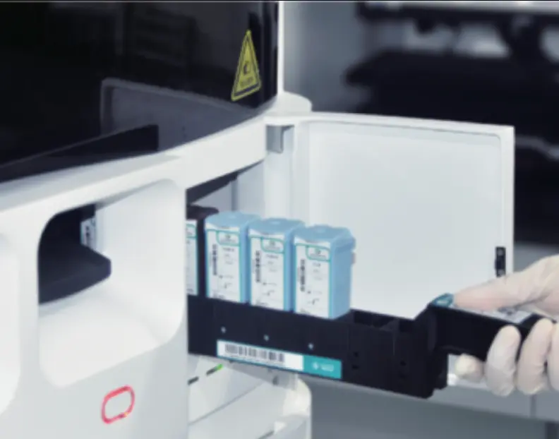
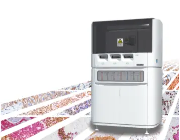
CNT 330: Automating Excellence in Multiplex Staining
To streamline workflows and achieve high-throughput staining capabilities, Celnovte provides the CNT 330 Full Automatic Multiplex IHC Stainer. This sophisticated instrument automates the entire mIHC staining process, minimizing human error, ensuring consistency, and freeing up researchers to focus on data analysis and scientific interpretation.
Celnovte’s Multiplex Immunofluorescence IHC User Guide: Guiding Researchers to Success
Celnovte is committed to supporting researchers in their mIHC endeavors, providing the Multiplex Immunofluorescence IHC User Guide. This comprehensive guide provides detailed protocols, troubleshooting tips, and valuable insights for optimizing mIHC experiments using Celnovte products, empowering researchers to achieve high-quality results and extract meaningful biological insights.
Automation and Standardization: Embracing Reproducibility and Efficiency
Automation plays a transformative role in mIHC, enhancing reproducibility, improving efficiency, and minimizing human error. Automated IHC staining platforms perform multiple staining steps with precision and consistency, ensuring uniformity across samples and reducing the variability inherent in manual staining procedures.
Interpretation and Analysis: Unveiling the Tapestry of Biological Insights
The real strength of mIHC lies in its capacity to derive biological understandings from the intricate staining patterns observed — requiring reliable imaging methods ᴛʜᴀᴛ ᴀʀᴇ sophisticated data analysis tools and diligent interpretation by experienced professionals.
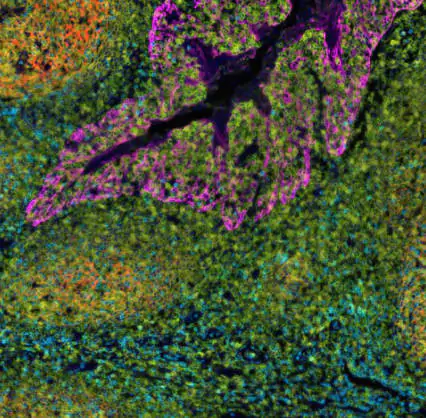
Multiplex Imaging: Capturing the Symphony of Colors
Specialized imaging platforms are essential for capturing and analyzing multi-channel fluorescent images generated in mIHC experiments. These systems utilize specific excitation and emission filters to distinguish between the different fluorophores used, enabling visualization of the individual target proteins and their spatial relationships within the tissue context. Confocal microscopy, with its ability to optically section through thick tissue samples, is particularly valuable in mIHC, allowing for the creation of high-resolution, three-dimensional images that reveal the intricate architecture of the tissue and the spatial distribution of biomarkers.
Data Analysis: Deciphering the Code of Multiplexed Signals
Specialized software applications are essential for examining mIHC data by measuring signal strength accurately and studying the relationships between various biomarkers in terms of their spatial positioning within tissues. These tools make it easier for researchers to analyze data objectively and consistently to gather details regarding protein levels and distribution patterns within the tissue environment and to explore potential interactions, among different cell types.
Conclusion
Multiplex immunohistochemistry (mIHC) shows potential to enhance our comprehension of intricate biological functions. It allows for the observation and examination of numerous biomarkers within their natural tissue setting offering unique perspectives on cell interactions signaling pathways and disease mechanisms. Nonetheless, just like any method’ obtaining consistent and significant outcomes with mIHC demands a focus on specifics fine-tuning and uniformity, across the complete experimental process.
In this blog post, we delved into the crucial elements that impact mIHC outcomes which include careful sample preparation methods along with efficient antigen retrieval procedures and strategic blocking techniques in addition, to choosing antibodies thoughtfully and ensuring their validation. All while selecting suitable detection systems and leveraging the advantages of automation and standardization.
Companies like Celnovte, with their commitment to innovation, quality, and customer support, play a crucial role in empowering researchers with the tools and expertise needed to navigate the complexities of mIHC and unlock its full potential. Their comprehensive portfolio of antibodies, reagents, instruments, and expert guidance enables researchers to overcome challenges, obtain reliable and high-quality data, and translate their findings into meaningful biological insights, ultimately contributing to advancements in diagnostics, therapeutics, and our understanding of human health.
