
-
-
SHOP

Multiplex IHC Insights: Distinguishing Between Immunohistochemistry and Immunocytochemistry
![]() 2024-12-10
2024-12-10
By admin
Immunohistochemistry (IHC) and immunocytochemistry (ICC) are methods in the fields of pathology and cellular biology. They both rely on antibodies to identify antigens within cells, but they vary greatly in their use and sample preparation.

Defining Immunohistochemistry
Immunohistochemistry is a method utilized for observing the location and distribution of proteins in tissue samples, commonly used in identifying cells seen in cancerous tissues within diagnostic pathology practices. By using antibodies that attach to particular antigens, IHC produces a detailed picture of protein positioning in the tissue structure. Typically employing chromogenic detection systems, like HRP-DAB, enables examination under a light microscope. This method proves to be highly beneficial in world clinical settings as it aids in identifying illnesses like EB virus infection using EBER in situ hybridization techniques.
Understanding Immunocytochemistry
Immunocytochemistry emphasizes examining cells rather than tissue samples and is a common practice for studying cultured cells or those extracted from bodily fluids. In ICC, the process involves adhering the cells to a slide and using antibodies to identify proteins in the cytoplasm or nucleus. This methodology plays a role in research scenarios requiring the detailed investigation of cell-specific protein expression independently of surrounding tissue frameworks.
How Do These Techniques Differ?
When it comes to identifying proteins using techniques, like IHC and ICC, that rely heavily on antibodies, the key differences lie in the types of samples they require, how they are prepared, their applications, and how they are typically used.
Sample Type and Preparation
The main difference between IHC and ICC is in the types of samples they use. The process involves staining tissue samples that are usually embedded in paraffin and thinly sectioned for analysis. This method maintains the tissues structure integrity which enables pathologists to evaluate not only protein expression but also its specific location, within the overall tissue framework.
In comparison to that method of analysis with ICC where they study cells that could come from either cultured systems or bodily fluids, the procedure requires placing these cells directly onto slides for examination which makes it easier but eliminates any details about the structure of the tissue.
Application and Usage
In settings, IHC is widely utilized for diagnostic purposes; specifically in cancer diagnosis where it detects tumor markers pivotal for treatment decisions and aids in improving surgical plans through rapid results during surgeries.
In contrast with that view expressed earlier in the paragraph about ICC’s usefulness manifests in research environments where discernment of cell-specific protein expression holds significance—it enables scientists to delve into cellular mechanisms at a detailed level, unhindered by surrounding tissues effects.
Solutions
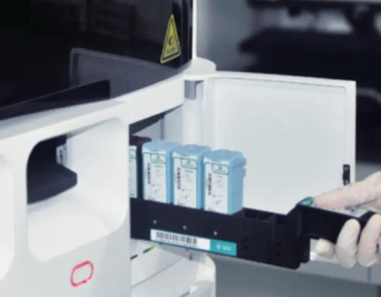
Looking for options to simplify multiplex IHC staining in labs? Delving into automation tools, such as the CNT330 Full Automatic Multiplex IHC Stainer, could be a great idea! These tools help streamline processes by automating staining procedures and maintaining top-notch outcomes across various samples.
For solutions that cater to individual laboratory requirements and challenges, make sure to explore the Solution Center at Celnovte. There you’ll discover assistance for seamlessly integrating advanced technologies into your everyday operations.
Why Is Multiplex IHC Important?
Multiplex immunohistochemistry (IHC), a method that allows for the simultaneous identification of multiple antigens in one tissue sample—an essential tool across diverse fields like cancer research, for deciphering intricate cell interactions and pinpointing various biomarkers impacting diagnostic and treatment approaches significantly.
Benefits of Multiplex IHC
Multiplex IHC offers several advantages over traditional single-marker staining techniques. It allows for the comprehensive analysis of cellular environments, providing insights into the spatial relationships between different cell types and proteins. This can lead to more accurate diagnoses and a better understanding of disease mechanisms. Additionally, multiplexing reduces the amount of tissue required for analysis, preserving valuable samples for further studies.
Innovations in Multiplex IHC Technology
Advances in multiplex IHC technology have transformed its use in both research and medical environments significantly. The introduction of automated systems such as the CNT330 Full Automatic Multiplex IHC Stainer has simplified intricate staining procedures, resulting in reliable and top-notch outcomes consistently. These developments not only boost productivity but also minimize mistakes made by humans, hence expanding the availability of multiplex IHC to labs across the globe.
What Are the Challenges Faced?
While multiplex IHC offers advantages, different issues must be resolved to make the most of its potential.
Technical Limitations
One major challenge in this aspect is fine-tuning antibody combinations to prevent any cross-reactivity or unintended binding that may affect result accuracy negatively. Managing signal overlaps between different dyes is crucial for distinguishing targets clearly and accurately. These technical challenges demand validation procedures and often call for specific tools and expertise to overcome them effectively.
Interpretation of Results
Analyzing outcomes from IHC can be tricky because of the large volume of data produced, requiring sophisticated image analysis tools and skilled individuals to differentiate actual signals from noise effectively based on multi-marker expression pattern evaluations, highlighting the importance of consistent procedures and rigorous quality checks for dependable interpretations.
Solutions
To tackle these obstacles successfully in labs like yours, it is advised to look into customized solutions provided at Celnovte’s Solution Center, for guidance and assistance in incorporating cutting-edge multiplex IHC technologies into your workflow to guarantee top-notch performance and precision in your evaluations.
Utilizing these cutting-edge solutions in your lab work can improve the capacity to perform advanced multiplex IHC tests that provide valuable information on disease progression and treatment effectiveness.
How Can These Challenges Be Addressed?
Navigating the intricacies of IHC requires careful planning to address its unique obstacles effectively, boosting precision and productivity through the utilization of sophisticated resources and practical strategies.
Solutions for Effective Implementation
To tackle the challenges of multiplex IHC effectively, labs need to take a thorough approach. They should concentrate on refining antibody sets to reduce any issues with cross-reactivity and non-specific binding. This includes conducting validation procedures to confirm that each antibody targets its intended molecule accurately without being influenced by other antibodies in the panel.
At Celnovte’s Solution Center, laboratories can use resources to simplify intricate staining procedures and enhance the accuracy of their findings.
Role of Advanced Kits and Tools
Using kits and tools is essential for addressing the difficulties linked to multiplex IHC testing techniques effectively. Automated systems such as the CNT330 Full Automatic Multiplex IHC Stainer are key in improving workflow efficiency by automating complex procedures and minimizing potential errors that guarantee consistent delivery of high-quality results.
Furthermore, coupling advanced image analysis software can assist in interpreting intricate datasets produced by multiplex IHC. This innovation enables precise distinction between authentic signals and background noise, enabling dependable evaluations of multiple markers’ expression levels.
What Is the Future Outlook?
The upcoming developments in multiplex IHC show promise for enhancing its integration in research and clinical settings.
Emerging Trends in Multiplex IHC
Current developments in IHC are geared towards enhancing the scope and sophistication of analysis capabilities in the field. Innovative technologies, like multiplexed platforms, are being engineered to simultaneously identify a larger array of antigens. These advanced platforms facilitate a thorough examination of cellular surroundings and yield a deeper understanding of disease processes.
Furthermore, there is an increasing focus on enhancing the precision and accuracy of detection systems. This includes the creation of fluorescent dyes that have limited signal interference, thus improving the sharpness and reliability of outcomes.
Integration with Other Technologies
The combination of IHC with other technologies is poised to transform its range of uses significantly. When this method is combined with proteomic analyses it can provide a comprehensive understanding of cellular processes paving the way for a closer connection, between molecular biology and pathology perspectives.
Moreover, progress in pathology is enabling smooth integration with multiplex IHC. High-quality digital imaging permits visualization and measurement of staining patterns, aiding in the formulation of stronger diagnostic assessments.
Laboratories can tap into the capabilities of multiplex IHC by tackling present obstacles, with creative approaches and adopting new trends effectively. This will advance knowledge and enhance clinical results by enabling more accurate diagnostic and treatment plans.
