IHC Testing Process: Essential Steps and Insights for Beginners

By admin
Exploring the Basics of IHC Testing
What is IHC?
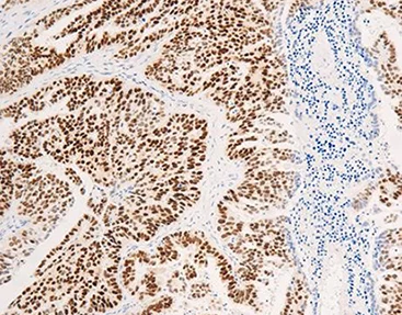
Immunohistochemistry (IHC) is a method used in laboratories to show where proteins are in tissue samples by using antibodies to find certain antigens present there. This helps researchers understand better the aspects of different illnesses, like cancer.
Importance of IHC in Medical Diagnosis
IHC plays a pivotal role in medical diagnostics by allowing pathologists to identify abnormal cells within a tissue sample. This technique aids in classifying tumors, determining their origin, and predicting patient prognosis. Celnovte has developed high-quality antibodies such as ER, PR, and HER2 that can clearly guide treatment plans.
Common Applications of IHC
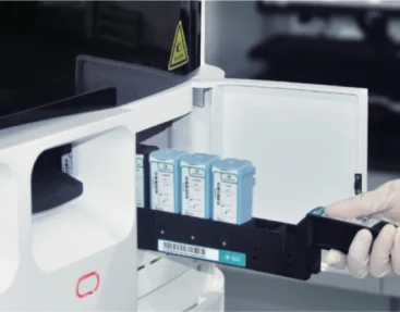
IHC is commonly applied in oncology to diagnose cancer types and subtypes. It is also used for identifying infectious agents and assessing the expression levels of various biomarkers related to disease progression and response to therapy. Celnovte’s fully automated IHC staining machine supports technologies like chromogenic in situ hybridization and multi-color IHC.
Preparing for an IHC Test
Sample Collection and Handling
The precision of an IHC test starts with gathering and managing samples. It’s important to use methods when collecting tissue samples to avoid contamination. After collection, it’s crucial to fix the samples to maintain the integrity of cellular structures.
Fixation Techniques
Ensuring fixation is essential for preserving tissue structure during processing tasks in the lab setting. Formaldehyde based fixatives are widely employed for this purpose; however, they necessitate handling due to their potentially harmful properties. Celnovte’s fixative has undergone scrutiny, in real world clinical scenarios to guarantee precise antibody staining outcomes.
Embedding and Sectioning Procedures
Upon completion of fixation process tissues are encased in paraffin blocks. Subsequently sliced thinly for analysis purposes. Precision is essential during this stage to maintain consistency, across sections ensuring uniform staining outcomes throughout.
Key Steps in the IHC Testing Process
Deparaffinization and Rehydration
Purpose of Deparaffinization
When tissue sections go through deparaffinization process remove the embedding medium so that antibodies can reach the antigens in the sample.
Rehydration Techniques
Rehydrating tissues entails dipping slides in lower concentrations of alcohol before rinsing with water restoring the tissue to its original state prior, to staining.
Antigen Retrieval Methods
Heat-Induced Epitope Retrieval (HIER)
HIER utilizes heat to reveal antigens that may have been hidden during fixation improving the effectiveness of antibody binding by restoring antigenicity.
Enzymatic Retrieval Techniques
Enzymatic extraction involves the use of enzymes, like proteinase K to break down proteins that cover antigen sites and make it easier for antibodies to reach them.
Blocking Non-Specific Binding Sites
Types of Blocking Agents Used
To prevent antibodies from binding blocking agents saturate potential sites on the tissue, such as normal serum or bovine serum albumin (BSA).
Evaluating Staining Patterns
|
Staining Pattern |
Interpretation |
|
Positive |
Target antigen is present; indicates successful staining |
|
Negative |
Absence of target antigen; may require protocol optimization |
|
Non-specific |
High background staining; suggests need for improved blocking |
The advanced multiplex fluorescence IHC technology developed by Celnovte enables in depth examination of tumor environments by revealing intricate cell interactions in tissues. Through the use of tyramide signal amplification (TSA), this method permits the simultaneous identification of various biomarkers on a single tissue sample without limitations, on antibody selection based on species.
This sophisticated approach enhances not only the sensitivity of target detection but also enables automated detection with great efficiency and reliability, making it easier to provide accurate cancer diagnoses, through improved imaging results.
Detection and Visualization in IHC
Primary Antibody Application
Selecting the Right Antibody
Selecting the right primary antibody is key for the accuracy and effectiveness of the IHC test process. It’s important that the primary antibody strongly binds to the desired antigen to guarantee detection. Celnovte’s proficiency in crafting top notch antibodies like ER, PR and HER2 guarantees outcomes, for treatment strategy guidance.
Incubation Conditions
The best conditions for breeding improve the interactions between antibodies and antigens by managing temperature and concentration while ensuring maximum binding efficiency and specificity are upheld.
Secondary Antibody Application and Detection Systems
Types of Detection Systems
Detection systems enhance the signal produced by the antibody attachment process often using enzyme linked secondary antibodies to trigger a color change reaction or using secondary antibodies labeled with fluorescent markers for immediate visualization purposes.
Enhancing Signal Visibility
Signal visibility can be improved using methods like tyramide signal amplification (TSA) which makes use of peroxidase to trigger a reaction, with a substance that bonds closely to proteins nearby and boosts the signal strength significantly. Celnovte multiplex fluorescence IHC technology utilizes TSA to achieve on site labeling of target proteins or nucleic acids.
Analyzing and Interpreting IHC Results
Evaluating Staining Patterns
|
Staining Pattern |
Interpretation |
|
Positive |
Indicates presence of target antigen; confirms successful staining |
|
Negative |
Absence of target antigen; may indicate need for protocol adjustment |
|
Non-specific |
High background staining; suggests a requirement for better blocking |
Celnotve’s groundbreaking multiplex fluorescence IHC technique offers an examination of the immune environments in tumors and enables a deep insight into the intricate interactions among cells, in tissues.
Troubleshooting Common Issues
Weak or No Staining
Insufficient or lacking coloration may occur due, to not retrieving the antigen using a low concentration of the primary antibody or improper incubation settings; adjusting these factors can improve the intensity of the coloration.
Background Noise Reduction
Optimizing the surrounding conditions can help minimize background noise when conducting experiments, with antibodies; another approach is to choose antibodies that are more targeted or to use thorough washing techniques to eliminate any unbound substances left behind after the process.
Celnovte’s Role in Advancing IHC Solutions
Innovative Products Offered by Celnovte
At Celnovte, they have a variety of cutting-edge products that are meant to enhance the results of IHC testing. With their fluorescence IHC kits, multiple biomarkers can be labeled simultaneously on one tissue section offering richer biological insights from limited samples.
Services Provided to Enhance IHC Testing Efficiency
Specializing in laboratory support services Celnovte offers solutions to streamline the IHC workflow process. Their automated systems guarantee outcomes, reducing the need for extensive manual handling and enhancing efficiency.
Commitment to Quality and Accuracy in Diagnostics
Operating under the belief that quality serves as the cornerstone of an enterprise, Celnovte remains dedicated to delivering top notch products and services with a focus on accuracy and precision in diagnostic applications.Their unwavering commitment has solidified their reputation as front-runners in propelling IHC technologies and fostering groundbreaking research initiatives on a global scale.
RELATED PRODUCTS


