IHC Detection Method Insights: Navigating Dual Techniques

By admin
In the realm of immunohistochemistry (IHC), the detection methods are pivotal for accurate analysis and diagnosis. These methods primarily revolve around the interaction between antigens and antibodies, followed by either enzymatic or fluorescent detection techniques.
The Role of Antigen-Antibody Reactions in IHC
Central to IHC is the antigen-antibody reaction, where specific antibodies bind to target antigens in tissue samples. This binding forms the foundation for detecting and visualizing proteins within cells. The specificity of this interaction ensures that only the target protein is highlighted, allowing for precise localization within tissue sections.
Enzymatic and Fluorescent Detection: Key Approaches
The subsequent step involves choosing between enzymatic and fluorescent detection methods. Enzymatic detection involves using enzymes attached to antibodies to trigger a color reaction that can be seen through a microscope lens. Differently, fluorescent detection utilizes antibodies labeled with fluorochrome that emit light when stimulated. This offers a method of visualization.
How Do Enzymatic Detection Methods Work in Practice?
Enzymatic detection methods are widely used due to their robustness and ease of interpretation. They convert an invisible antigen-antibody interaction into a visible signal through an enzymatic reaction.
Mechanism Behind Enzymatic Labeling
Enzymatic labeling involves conjugating enzymes like horseradish peroxidase (HRP) to secondary antibodies. Upon binding to the primary antibody-antigen complex, a substrate is added, leading to a color change that can be observed under a light microscope. This method is exemplified by technologies like Super-ISH super chromogenic in situ hybridization, which uses HRP-DAB systems for clear visualization.
Advantages and Limitations of Enzymatic Techniques
Enzymatic techniques offer several advantages, including high sensitivity and permanent staining results that are easy to interpret using standard microscopy. However, they can be limited by factors such as background staining or cross-reactivity if not properly optimized.
For those seeking advanced solutions tailored to specific needs in IHC detection methods, visiting Celnovte’s Solution Center could provide valuable insights and options.
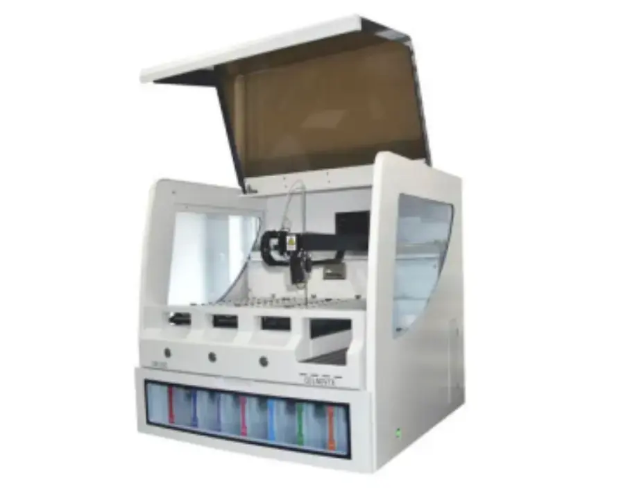
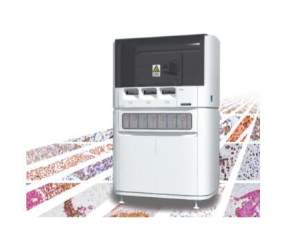
In conclusion, understanding IHC detection methods requires navigating through antigen-antibody interactions and selecting appropriate visualization techniques. Whether opting for enzymatic or fluorescent approaches, each has its distinct set of benefits and challenges that must be considered in clinical and research settings. For automated solutions that enhance efficiency in IHC processes, exploring products like CNT330 Full Automatic Multiplex IHC Stainer can significantly streamline workflows.
Why Choose Fluorescent Detection Methods?
In the landscape of IHC detection methods, fluorescent techniques offer unique advantages that make them appealing for various applications. These methods utilize fluorochrome-labeled antibodies to provide a distinct visualization approach.
The Science Behind Fluorescent Labeling
Using labeling entails attaching fluorescent dyes to antibodies that emit light upon stimulation by particular wavelengths. This emission enables the visualization of proteins in tissues, through fluorescence microscopy. The effectiveness of this method lies in its capacity to detect targets simultaneously by employing various fluorochromes that do not interfere with each other.
Pros and Cons of Fluorescent Techniques
Fluorescent detection techniques are well known for their accuracy and ability to analyze antigens at once in a sample with a high level of sensitivity and efficiency. This is especially useful in situations where different markers need to be studied in complex tissue settings. However, the equipment and skills needed for these methods can be specialized. There might be issues with the fluorescence signal weakening over time making it difficult, for long-term research projects.
How Can You Optimize Your IHC Protocols?
Optimizing IHC protocols is crucial to achieving reliable and reproducible results. Several factors influence the selection of appropriate IHC detection methods and the overall accuracy of the analysis.
Factors Influencing IHC Method Selection
Selecting the right IHC method depends on various factors, including the nature of the tissue sample, the target antigen’s abundance, and the desired sensitivity level. Consideration must also be given to available equipment and expertise within your laboratory setting. Tailoring these aspects ensures that chosen methods align with specific research or clinical objectives.
Enhancing Accuracy with Advanced Equipment
Utilizing advanced equipment can significantly enhance the accuracy and efficiency of IHC processes. Automated systems like CNT330 Full Automatic Multiplex IHC Stainer streamline workflows by reducing manual intervention, minimizing variability, and ensuring consistent staining quality across samples.
For further insights into optimizing your protocols with tailored solutions, exploring resources such as Celnovte’s Solution Center can provide valuable guidance and support.
In summary, navigating through IHC detection methods involves understanding both enzymatic and fluorescent approaches, each offering distinct benefits suited to different applications. By carefully selecting methodologies based on specific requirements and leveraging advanced technologies, you can enhance both efficiency and accuracy in your immunohistochemical analyses.
Solutions for Common Challenges in IHC Detection
Navigating through the complexities of IHC detection methods often presents challenges, but solutions are available to enhance accuracy and reliability.
Addressing Issues with Background Staining
Background staining can obscure results and lead to misinterpretation. To address this, consider optimizing antibody concentrations and ensuring thorough washing steps during the protocol. Employing blocking agents such as serum or specific proteins can also minimize non-specific binding, thus reducing background noise. For specialized assistance in refining your protocols, Celnovte’s Solution Center offers comprehensive resources and expert guidance.
Optimizing Signal-to-Noise Ratio
A high signal-to-noise ratio is crucial for clear visualization of target antigens. This can be achieved by selecting highly specific antibodies and using amplification systems that enhance signal strength without increasing background. Additionally, adjusting incubation times and temperatures can further refine the balance between signal clarity and background interference.
How Is Celnovte Pioneering Innovations in IHC?
Celnovte stands at the forefront of innovation in IHC detection methods, offering cutting-edge solutions that streamline processes and improve outcomes.
Cutting-edge Products from Celnovte
Celnovte’s product line includes state-of-the-art technologies designed to enhance IHC workflows. The CNT330 Full Automatic Multiplex IHC Stainer exemplifies this innovation by automating complex staining procedures, thereby reducing manual errors and increasing throughput. This equipment ensures consistent quality across samples, making it an invaluable asset for laboratories aiming to boost efficiency and accuracy.
In conclusion, addressing common challenges in IHC detection involves strategic adjustments to protocols and leveraging advanced technologies. By utilizing resources like Celnovte’s innovative products and solutions center, you can significantly enhance the quality and reliability of your immunohistochemical analyses.
RELATED PRODUCTS


