
-
-
SHOP

How Do mIHC Staining and Biopsy Techniques Differ?
![]() 2024-12-19
2024-12-19
By admin
When evaluating tissue samples, professionals often choose between multiplex immunohistochemistry (mIHC) staining and biopsy techniques. Each method offers unique insights, but understanding their differences is crucial for accurate diagnosis and research.

The Basics of mIHC Staining
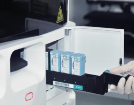
Multiplex immunohistochemistry (mIHC) is a method that enables the concurrent identification of multiple biomarkers in a single tissue section offering significant advantages in areas such, as cancer research and immunology by providing a detailed insight into intricate biological interactions all at once. Techniques such as the CNT-300 Automatic IHC Slide Stainer facilitate this process by automating the staining, ensuring precision and consistency.
The Fundamentals of Biopsy Procedures
During a procedure samples of tissue are taken from the body for disease examination. Unlike mIHC which looks at cellular markers, biopsy offers a more comprehensive view enabling pathologists to study the tissue’s structure and form. This technique is critical for diagnosing conditions like cancer, where physical characteristics of cells can indicate malignancy.
What Are the Advantages of mIHC Staining?
Given its ability to detect multiple biomarkers simultaneously, mIHC staining presents several advantages over traditional biopsy methods.
Enhanced Precision and Accuracy
mIHC staining enhances diagnostic precision by allowing detailed examination at a molecular level. It provides clearer insights into cellular processes and interactions, which are essential for accurate disease characterization. This precision is particularly evident when comparing signal intensities across different channels, where CNT’s multiplex solutions have shown superior performance in delivering strong signal-to-noise ratios.
Advanced Diagnostic Capabilities
The advanced capabilities of mIHC staining extend beyond mere detection. By offering deeper insights into biological systems, it aids in understanding disease mechanisms more comprehensively. Researchers can leverage these insights to develop targeted therapies and improve patient outcomes significantly. For those looking to integrate these advanced techniques seamlessly into existing workflows, Celnovte’s solutions offer adaptable options that ensure minimal disruption while maximizing diagnostic potential.
In summary, both mIHC staining and biopsy techniques play pivotal roles in medical diagnostics and research. While biopsies provide essential morphological information necessary for initial diagnoses, mIHC offers enhanced molecular insights crucial for comprehensive disease analysis and treatment planning. By incorporating tools like CNT-300, professionals can achieve greater accuracy and efficiency in their diagnostic processes.
Why Is Biopsy Still a Preferred Method in Certain Cases?
Despite the advancements in mIHC staining, biopsy remains a preferred method in specific scenarios due to its unique advantages.
Comprehensive Tissue Analysis
Examining tissue morphology through biopsy is crucial in diagnosing diseases such as cancer as it offers a view of cellular structure and abnormalities that cannot be detected solely through molecular analysis. Obtaining insights into morphology from biopsies plays a key role, in detecting malignancies and various pathological conditions.
Established Clinical Protocols
Biopsies have long been integrated into clinical protocols and are widely accepted as a standard diagnostic procedure. Their reliability and established guidelines make them a trusted choice for clinicians. These protocols ensure consistent results across various medical settings, providing a solid foundation for disease diagnosis and treatment planning.
Can mIHC Staining Replace Traditional Biopsy Methods?
As mIHC staining gains popularity, questions arise about its potential to replace traditional biopsy methods.
Comparative Effectiveness
Though mIHC staining can give us molecular information that is quite advanced; it is seen more as a supplement rather than a substitute for biopsies in the medical field. Being able to identify biomarkers all at once can give us significant insights into how cells interact and the mechanisms behind diseases. Nevertheless, for medical conditions and diagnoses; the thorough examination of tissues, through biopsies continues to hold its crucial place and cannot be replaced by any other method. Both techniques serve distinct roles in medical diagnostics, with mIHC enhancing the depth of analysis when used alongside traditional methods.
Limitations and Challenges
Despite its advantages, mIHC staining faces certain limitations. The intricacy of the method demands tools and know-how that might pose a challenge for use in certain medical environments or settings to implement effectively. Moreover, even though mIMC offers, in-depth information it may fall short of capturing the complete intricacies of tissue structure needed for certain diagnoses. Therefore, integrating both mIHC and biopsy techniques ensures a more holistic approach to patient care.
In summary, although mIHC staining brings progress in molecular diagnostics conventional biopsy approaches still maintain their position thanks to their thorough tissue examination abilities and well-established clinical procedures. When combining both methods medical professionals can reach precise diagnoses and enhance patient results effectively. For those interested in exploring innovative solutions that integrate these methodologies seamlessly into existing workflows, Celnovte’s solutions provide adaptable options tailored to meet diverse diagnostic needs.
How Do You Choose Between mIHC Staining and Biopsy?
When deciding between mIHC staining and biopsy, several factors come into play. Each technique has its strengths and limitations, making the decision process complex yet critical for achieving accurate diagnostic outcomes.
Factors Influencing Decision-Making
The decision between opting for mIHC staining or a biopsy typically hinges on the clinical situation and the type of information needed at hand. For example, if delving into a molecular analysis becomes imperative to grasp the core disease mechanisms in play mIHC staining could be the preferable choice. This method enables the identification of multiple biomarkers offering a thorough perspective, on cellular interactions.
Alternatively in cases where examining the structure of tissues is essential for diagnosis. Like identifying structures in tissues. A biopsy might be a better option to consider. The detailed analysis of tissue architecture that biopsies allow for is invaluable, in situations especially when it comes to diagnosing cancerous conditions.
Role of Medical Expertise
The expertise of medical professionals also plays a significant role in determining the appropriate technique. Experienced pathologists can leverage their knowledge to interpret complex data from both mIHC staining and biopsy procedures effectively. Their ability to integrate molecular insights from mIHC with morphological data from biopsies enhances diagnostic accuracy and informs treatment decisions.
Incorporating advanced tools like the CNT-300 Automatic IHC Slide Stainer can streamline processes, ensuring that both techniques are utilized optimally within clinical workflows. By doing so, healthcare providers can achieve a balanced approach that maximizes diagnostic potential.
Solutions for Integrating mIHC Staining and Biopsy
For those seeking to integrate both mIHC staining and biopsy techniques seamlessly into their diagnostic workflows, Celnovte’s solutions offer innovative options. These solutions are designed to enhance compatibility between methods, ensuring minimal disruption while maximizing efficiency and accuracy.
By adopting these integrated solutions, healthcare professionals can leverage the strengths of both techniques, achieving comprehensive insights that drive better patient outcomes. Whether utilizing automated platforms or manual methods, Celnovte provides adaptable options tailored to meet diverse clinical needs.
In conclusion, choosing between mIHC staining and biopsy involves careful consideration of various factors including clinical requirements and medical expertise. By understanding the unique advantages each method offers and integrating them effectively into existing workflows with advanced solutions like those from Celnovte, healthcare providers can ensure precise diagnostics and improved patient care.
What Solutions Are Available for Integrating Both Techniques?
Combining both mHIC staining and biopsy methods, in practice can greatly improve diagnostic abilities as it allows healthcare providers to gain a deeper insight into intricate diseases.
Combined Diagnostic Approaches
A combined diagnostic approach involves using mIHC staining and biopsy techniques in tandem to capitalize on their unique advantages. This strategy allows for a more holistic analysis by integrating molecular insights from mIHC with the morphological data obtained from biopsies. The simultaneous detection of multiple biomarkers through mIHC provides detailed information about cellular interactions, while biopsies offer an in-depth examination of tissue architecture. Together, they provide a multi-faceted view that enhances diagnostic precision and informs treatment decisions.
Technological Advancements
Technological advancements play a crucial role in facilitating the integration of mIHC staining and biopsy methods. Automated platforms like the CNT-300 Automatic IHC Slide Stainer streamline the staining process, ensuring consistency and accuracy across samples. These innovations reduce manual intervention, minimize errors, and increase throughput, making it easier to incorporate both techniques into routine practice.
For those seeking to optimize their diagnostic workflows further, Celnovte’s solutions provide adaptable options that enhance compatibility between methods. By adopting these advanced solutions, healthcare providers can seamlessly integrate mIHC staining and biopsy procedures, achieving comprehensive insights that drive better patient outcomes.
In summary, integrating mIHC staining and biopsy techniques requires careful consideration of combined diagnostic approaches and technological advancements. By leveraging these strategies effectively, healthcare professionals can maximize the diagnostic potential of both methods, ensuring precise diagnostics and improved patient care.
What Does the Future Hold for mIHC Staining and Biopsy Techniques?
Emerging Trends in Medical Diagnostics
The field of testing is changing quickly as mIHC staining and biopsy methods take center stage in these advancements. As new technologies develop some emerging trends are set to transform the way illnesses are detected and managed. One notable trend is the growing incorporation of artificial intelligence (AI) in diagnostic procedures. Through AI algorithms that analyze data sets, from mIHC staining new possibilities arise for understanding disease processes and making accurate diagnoses.
Moreover, there is a growing emphasis on personalized medicine, where diagnostic techniques are tailored to individual patient profiles. This approach leverages the detailed molecular information provided by mIHC staining to develop targeted therapies that improve patient outcomes. As these trends continue to gain traction, mIHC staining and biopsy techniques will play an increasingly vital role in advancing medical diagnostics.
Ongoing Research and Development
Research and development play a role in improving the effectiveness of mIHC staining and biopsy methods. Scientists are constantly looking into biomarkers that mIHC testing can identify to broaden its use in fields, like oncology and immunology. Additionally, the progress made in imaging technologies is enhancing the precision and reliability of both mIHC staining and biopsy assessments.
Joint research projects are also driving creativity by uniting specialists from different fields to tackle intricate diagnostic issues together. This work is paving the path, for approaches that combine both immunohistochemical staining and biopsy techniques to provide a thorough understanding of disease pathology.
For those interested in staying at the cutting edge of diagnostic technology, Celnovte’s solutions provide innovative options that enhance compatibility between traditional and advanced methods. By embracing these developments, healthcare professionals can ensure they remain at the forefront of medical diagnostics.
In summary, the outlook for mIHC staining and biopsy methods appears promising, as new developments and continuous studies are leading to progress in medical testing. By utilizing these advancements medical professionals can attain more precise diagnoses and enhance patient care results.