
-
-
SHOP

How Cytology Stainers Revolutionize Cellular Analysis
![]() 2024-08-13
2024-08-13
By admin

The Importance of Advanced Cytology Techniques
Cytology stainer is an essential method in cellular analysis, aimed at improving the visualization of cells and their components when viewed under a microscope. This technique involves the use of specific stains that attach to cellular structures, enabling researchers and clinicians to differentiate and identify various cell types, abnormalities, and pathological conditions. By grasping the principles of cytology stains, professionals can accurately interpret the morphological features of cells, which is vital for precise diagnosis and research.
Principles of Cytological Stains
The principles of cytological stains involve the interaction between dyes and cellular structures. These dyes exhibit affinity towards certain cellular components, such as nucleic acids and proteins, enabling their differentiation based on color changes. Common stains like Hematoxylin and Eosin (H&E) selectively color cell nuclei and cytoplasm, respectively, providing a contrast that facilitates the identification of cellular details. Advanced techniques, such as chromogenic in situ hybridization, use enzyme-linked reactions to detect specific RNA sequences, offering precise molecular pathology insights.
Historical Development in Cytology Staining Methods
The evolution of cytology staining methods has significantly contributed to the field of cellular analysis. Early techniques relied on basic dyes and manual staining procedures, which were labor-intensive and prone to inconsistencies. With technological advancements, automated staining machines and more sophisticated dyes have emerged, enhancing the accuracy and efficiency of staining processes. Innovations like multi-color immunohistochemistry and double staining for biomarkers such as p16/Ki-67 have revolutionized the detection and study of complex pathological conditions, particularly in cancer diagnostics and research.
The Role of Cytology Stainers in Cellular Analysis
Enhancing Diagnostic Accuracy
Cytology stainers play a critical role in improving diagnostic accuracy by ensuring consistent and precise staining of cellular samples. These automated systems minimize human error and variability, providing reproducible results that are essential for reliable diagnosis. For instance, cytology stainers used in the detection of cervical cancer rely on color interpretation of biomarker stains like p16 and Ki-67, which are crucial for identifying precancerous lesions and reducing misdiagnosis.
Accelerating Workflow Efficiency
Efficiency stands out as a major benefit of employing cytology stainers in cellular analysis. These automated devices drastically cut down the time needed for sample preparation and staining, thus facilitating higher throughput in both clinical and research settings. With the ability to process multiple samples at once, these stainers enhance workflow efficiency, allowing pathologists and researchers to concentrate on analysis and interpretation. For instance, fully automated immunohistochemistry stainers can finalize staining processes in just 2.5 hours, meeting the rigorous demands of contemporary diagnostic practices.
Improving Sample Preservation
Sample preservation is another crucial benefit of utilizing cytology stainers. These machines apply stains evenly and consistently, ensuring that cellular samples retain their integrity and quality throughout the analysis process. Properly stained samples are less susceptible to degradation, which is vital for obtaining accurate and reliable results, especially in long-term studies and archival purposes. Enhanced sample preservation also facilitates secondary testing and re-evaluation, which are often necessary in complex diagnostic scenarios.
Technological Innovations in Modern Cytology Stainers
About Celnovte
Celnovte is a leader in the development and production of advanced cytology staining solutions, committed to enhancing the capabilities of cellular analysis. Their innovative technologies have set new standards in the field, providing robust and efficient tools for both clinical diagnostics and research applications. Celnovte’s dedication to quality and precision is evident in their cutting-edge products, which are designed to meet the highest standards of accuracy and reliability.
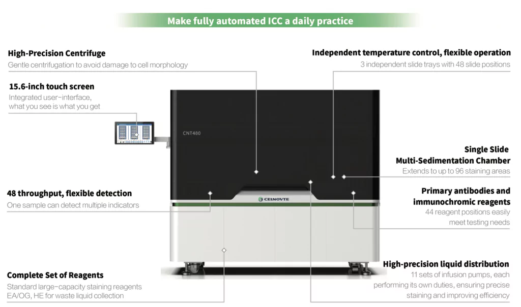
CNT 480 Fully Automatic Liquid-Based Cytology & Immunocytochemistry Stainer
The CNT 480 Fully Automatic Liquid-Based Cytology & Immunocytochemistry Stainer is a revolutionary tool in the field of cytology and immunocytochemistry. This advanced stainer automates the staining process for slides prepared using liquid-based cytology techniques, significantly enhancing efficiency and reducing processing time. Its automation not only streamlines the workflow but also ensures consistent staining quality, which is crucial for accurate diagnosis.
One of the key strengths of the CNT480 is its versatility. It is capable of staining a diverse range of cytology samples, including Pap smears, cervical exfoliated cells, endometrial cells, pleural effusion, ascites, pericardial effusion, urinary exfoliated cells, bone marrow cells, and other puncture cells. This versatility makes it an invaluable asset in any laboratory dealing with a wide variety of cytology specimens. Furthermore, the CNT480 has been proven to detect various markers alongside Pap on the same slide, such as CK7, TTF-1, CA125, CD45, CK5&6, CK-pan, and EMA, demonstrating its outstanding staining quality and reliability.
Efficiency is another hallmark of the CNT480. With its automation, the stainer allows for higher throughput, enabling the processing of more slides in a shorter period. Its design features 3 independent slide trays, each with 48 slide positions, along with 44 reagent positions. Each slide can accommodate 2 chambers, further increasing its capacity. Moreover, the CNT480 incorporates gentle centrifugation to avoid damaging cell morphology and utilizes 11 sets of infusion pumps for high-precision liquid dispensing. It also provides independent temperature control for each slide, ensuring precision and consistency in staining operations. These features combined make the CNT480 a powerful and efficient tool for cytology and immunocytochemistry laboratories.
Applications of Cytology Stainers in Medical and Research Fields
Use in Clinical Diagnostics
In clinical diagnostics, cytology stainers play a crucial role in detecting and analyzing various diseases. By delivering consistent and high-quality staining, these machines allow pathologists to accurately identify cellular abnormalities and pathological conditions. This is especially significant in cancer screening and diagnosis, where precise interpretation of cell morphology and biomarkers is vital. For instance, the use of p16/Ki-67 double staining has become essential in cervical cancer screening, providing reliable results that assist in early detection and effective treatment planning.
Application in Cancer Research
Cancer research greatly benefits from the capabilities of modern cytology stainers. These devices facilitate the study of tumor biology by enabling detailed visualization of cancerous cells and their interaction with surrounding tissues. Advanced staining techniques, such as multi-color immunohistochemistry, allow researchers to observe multiple antigen targets on a single slide, providing insights into the tumor microenvironment and aiding in the identification of potential therapeutic targets. This comprehensive approach enhances the understanding of cancer progression and the development of novel treatment strategies.
Role in Infectious Disease Studies
Cytology stainers also play a vital role in the study of infectious diseases. By accurately staining and preserving microbial samples, these machines help in the identification and characterization of pathogens. Techniques such as chromogenic in situ hybridization are particularly useful in detecting specific RNA sequences of infectious agents. This has practical implications for diagnosing and understanding infections, such as EBV, where in situ hybridization has become a gold standard in determining EBV infection in tissue samples. Accurate staining is crucial for advancing research in infectious diseases, leading to improved diagnostic methods and therapeutic approaches.
Optimizing Lab Performance with Advanced Cytology Stainers
Quality Control Measures
Implementing stringent quality control measures is pivotal in optimizing lab performance with advanced cytology stainers. Regular monitoring and validation of staining protocols ensure that the results are consistent and reliable. This includes calibrating the stainers to maintain the accuracy of dye application and periodically checking the quality of stains and reagents. Additionally, established benchmarks and controls for staining results can help detect any deviations or anomalies promptly. The integration of automated systems in cytology stainers also aids in minimizing human error, thus enhancing the reproducibility of results across different samples and experiments.
Training and User Proficiency
The effective utilization of cytology stainers significantly hinges on the proper training and proficiency of lab personnel. It is imperative to implement comprehensive training programs for technicians and pathologists, aimed at acquainting them with the operation of these sophisticated machines and the interpretation of stained samples. Incorporating hands-on sessions and providing instructional materials can significantly enhance their grasp of specific staining techniques and the resolution of common issues. Conducting proficiency testing, wherein users regularly assess their skills and knowledge, can ensure that they maintain a high level of competence in using the stainers both efficiently and accurately. Additionally, continuous education and updates on emerging technologies and protocols are essential to keep users abreast of the latest advancements in cytology staining.
Maintenance and Calibration Protocols
Routine maintenance and precise calibration of cytology stainers are fundamental for sustained optimal performance. Establishing a regular maintenance schedule helps prevent malfunctions and extend the lifespan of the equipment. This includes cleaning the machine components, replacing worn-out parts, and ensuring that the reagents are within their usable lifespan. Calibration protocols involve adjusting the stainer settings to achieve the desired staining intensity and consistency. Using standardized calibration slides and reagents can aid in maintaining the accuracy and reliability of the staining results. Accurate record-keeping of maintenance and calibration activities is also critical for tracking performance and compliance with regulatory standards.
Conclusion
The integration of advanced cytology stainers has undeniably revolutionized cellular analysis, offering significant improvements in diagnostic accuracy, workflow efficiency, and sample preservation. By embracing technological innovations such as those offered by Celnovte‘s CNT 480, medical and research laboratories can achieve unparalleled consistency and quality in their staining processes. The applications of cytology stainers extend across various medical fields, from enhancing clinical diagnostics and cancer research to improving the study of infectious diseases. Through stringent quality control measures, comprehensive training programs, and routine maintenance and calibration protocols, laboratories can fully harness the capabilities of these sophisticated machines. Consequently, the advancements in cytology staining technology continue to drive forward the boundaries of cellular analysis, fostering better patient outcomes and groundbreaking research discoveries in the biomedical field.
