
-
-
SHOP

Five Steps for Great IHC Images
![]() 2024-11-28
2024-11-28
By admin
In the field of biological studies, it’s vital to visualize the tiny world inside tissues. Think about pinpoint accuracy in detecting proteins within a cell to discovering a small beacon in a vast cellular terrain. That’s the magic of immunohistochemistry (IHC). This method serves as a spotlight on the level of highlighting proteins in tissue samples using antibodies. These antibodies act, like directed missiles attaching to their protein targets (antigens) allowing us to see exactly where and how plentiful they are situated. Exploring the workings of cells and diseases is akin to wielding a detective’s toolbox through IHC analysis. Shedding light on protein distribution and localization is crucial, for unraveling cellular mysteries and disease intricacies alike.
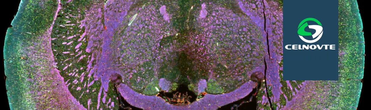
mIHC and the Power of Multiplexing
Picture this. Envision a scenario where you can observe proteins at once with each one marked in a different color on what looks like a vibrant and intricate cellular artwork piece. Enter the realm of multiplex immunohistochemistry ( mIHC ). With the ability to identify protein targets on a single tissue sample at the same time mCIH is revolutionizing the field of immunohistochemistry. This cutting-edge technique is transforming research efforts in fields such as cancer immunology by offering an understanding of the intricate interactions between cells and proteins, in the tumor microenvironment.
Celnovte Biotech is a leading provider of innovative mIHC solutions, offering researchers cutting-edge tools to unlock the potential of multiplexing. Explore Celnovte’s Solution to discover how their advanced technologies can elevate your research.
5 Steps to Publication-Quality IHC Images
Creating IHC images that are of high quality for publication involves paying close attention to every detail during the staining process—a bit like crafting a symphony where each stage plays a role, in the overall masterpieces outcome! Let me walk you through the five steps to achieve this art form;
Step 1: Sample Preparation
Tissue Preservation
The journey to great IHC images begins with the careful preservation of the tissue sample. It’s similar to setting the groundwork for a construction project – having a solid base is crucial for a sturdy structure to be built upon correctly. Ensuring preservation of tissues is key, in maintaining the integrity of cellular structures and protein components intact to prevent any degradation or signal loss. Two common methods are used:
Freezing: This technique includes freezing tissue samples using liquid nitrogen to stop enzymatic processes and maintain cellular integrity intact. It is common to use sections when identifying delicate antigens that could be harmed during chemical fixation.
Paraffin Embedding: The procedure involves using a chemical fixative such, as formalin to treat the tissue before embedding it in paraffin wax to preserve and store tissue samples term.
Choosing the right preservation method depends on the specific research goals and the nature of the target proteins.
Step 2: Antigen Retrieval
Epitope Unmasking
Picture a locked treasure chest with its secrets concealed from sight. Antigens are the focus of our antibodies. Can occasionally be obscured or tucked away in the tissue as a result of fixation or processing techniques. Unveiling these antigens is essential through antigen retrieval—it reveals the epitopes (binding sites) of our target proteins for antibody binding. This process is critical, for boosting staining intensity and obtaining precise signals. Two common methods are used:
Heat-Induced Epitope Retrieval (HIER): This technique involves using heat from devices like pressure cookers or microwaves to separate bonds that cover epitopes in tissue samples. The tissue slices are soaked in a retrieval solution such as citrate buffer (at pH 6.0). Tris-EDTA buffer (at pH 9.0). Then heated to a specific temperature, for a set amount of time.
Protease-Induced Epitope Retrieval (PIER): This method utilizes enzymes like proteinase K or trypsin to degrade proteins that could obstruct sites, on cells or molecules known as epitopes. Optimal handling is essential when using this approach to prevent breakdown and harm to tissue structure.
The choice of antigen retrieval method depends on the nature of the target protein and the fixation method used.
Step 3: Block
Minimize Non-Specific Signals
Imagine a detective trying to identify a suspect in a crowded room filled with distractions. Non-specific binding is like those distractions in IHC, creating background noise that can obscure the true signal. Antibodies, despite their specificity, can sometimes bind to unintended targets in the tissue, leading to false-positive results. Blocking is a crucial step in IHC to minimize this background noise, ensuring a clear and specific signal. It’s like silencing the unwanted whispers in a room to hear the main conversation.
Several blocking reagents are used to address specific sources of background:
Protein Blockers: These substances like serum albumin (BSA) or ordinary serum interact with general protein binding spots in the tissue to block antibodies, from binding to those areas.
Endogenous Enzyme Blockers: Tissues often contain endogenous enzymes, such as peroxidase or alkaline phosphatase, that can interfere with the detection system used in IHC. Specific blockers are used to inhibit these enzymes, reducing background staining.
Avidin/Biotin Blocking Reagents: When avidin biotin detection systems are utilized in experiments or tests reagents are employed to obstruct the biotin and avidin attachment sites. This action helps in averting the binding of the detection substances.
Careful selection and optimization of blocking reagents are essential for achieving clean and specific IHC staining.
Step 4: Detect
Target Antigen with Antibodies
This step is like the detective finally meeting the suspect face-to-face. The key player in this stage is the antibody tailored to detect our target protein effectively and accurately. The selection of the antibody holds significant importance and demands thoughtful evaluation of various factors such, as;
Specificity: The antibody must bind specifically to the target protein and not to other proteins in the tissue.
Sensitivity: The antibody should be able to detect the target protein even at low abundance levels.
Application: The antibody should be validated for use in the specific IHC protocol being used, as antibodies can perform differently in different applications.
Detection methods can be direct or indirect:
Direct Detection: In this method, the primary antibody is directly labeled with a detection system, such as a fluorophore or an enzyme. This method is simpler and faster but might offer lower sensitivity.
Indirect Detection: The process involves two steps; first an unmodified main antibody binds to the target. Then a labeled secondary antibody identifies the main one. This method offers higher sensitivity due to signal amplification.
Celnovte Biotech offers an extensive portfolio of high-quality primary antibodies for IHC applications, including their innovative MicroStacker™ Detection Systems, designed for superior sensitivity and specificity. Check out the CNT330 Full Automatic Multiplex IHC Stainer to experience the cutting-edge of automated IHC staining.
Step 5: Visualize
Capture Tissue Images
The last stage of IHC involves capturing the beauty of the stained tissue slice and showcasing the detailed molecular patterns we have uncovered through our work diligently done. This phase includes choosing the imaging method and mounting material to protect and present the stained sample effectively.
Imaging Modalities:
Light Microscopy: Used for visualizing chromogenic IHC stains, where the target proteins are labeled with colored substrates.
Fluorescence Microscopy: Used for visualizing fluorescent IHC stains, where the target proteins are labeled with fluorophores. This method offers higher sensitivity and the ability to multiplex, detecting multiple targets simultaneously.
Confocal Microscopy: An advanced form of fluorescence microscopy that provides optical sectioning, allowing for the creation of 3D images of the stained tissue.
Mounting Media:
Aqueous Mounting Media: Water-based media used for mounting chromogenic IHC stains.
Antifade Mounting Media: Special media used for mounting fluorescent IHC stains. These media contain chemicals that prevent the fading of fluorophores, preserving the signal for long-term imaging.
Selecting the imaging technique and mounting material is essential, for capturing top-notch IHC photographs that faithfully depict the staining outcomes.
Tips and Tricks for Optimizing IHC Staining
Optimization of IHC staining requires a balance of various elements to attain precise and consistent outcomes – akin to tuning a musical instrument, to achieving flawless harmony. Here are some valuable tips from the sources:
Ensure High-Quality Sections:
Section Thickness: Thinner sections, typically 4-5 micrometers, provide better resolution and allow for more even penetration of reagents.
Thorough Drying: Adequate drying of sections onto slides is crucial to prevent tissue detachment during staining.
Use of Charged Slides: Positively charged slides enhance tissue adhesion, minimizing section loss during staining.
Avoid Section Adhesion Problems:
Avoid Protein-Based Adhesives: Especially on charged slides, these adhesives can interfere with tissue adhesion, leading to uneven staining.
Prevent Concentration Gradients:
Adequate Reagent Volume: Use sufficient reagent volume to ensure even coverage of the tissue section.
Gentle Agitation: During the incubation process it can be helpful to gently agitate to ensure that the reagents are evenly distributed and avoid the formation of concentration gradients.
Optimize Nuclear Counterstaining:
Counterstain Intensity: Adjust the concentration and incubation time of the nuclear counterstain, such as hematoxylin, to achieve optimal contrast without masking weak specific staining.
Use Appropriate Controls:
Positive Controls: Tissues known to express the target protein should be included to ensure the staining protocol is working correctly.
Negative Controls: Tissues lacking the target protein or samples processed without the primary antibody serve to assess background staining and non-specific binding.
Consider Using TSA for Increased Sensitivity and Multiplexing:
TSA Technology: Employ tyramide signal amplification to enhance signal intensity, especially for low-abundance targets.
Multiplex Staining: Design panels using multiple primary antibodies and distinct fluorophores to visualize multiple targets simultaneously.
Conclusion
IHC, and especially with the developments in mIHC, is an essential cornerstone in research and diagnostics, allowing a view into the complex molecular microcosm of tissues. Publication-grade IHC images require meticulous attention to detail throughout the staining procedure, from the step of sample preparation to visualization. With a grasp of the principles of IHC and the implementation of best practices, researchers can tap into the complete potential of this potent technique, driving new findings and patient care forward.
Learning IHC is similar to learning any other skill; you must practice, be patient, and be open to learning and embracing new technology. Businesses such as Celnovte Biotech are at the forefront, offering innovative solutions to assist researchers on their journey to unveil the mysteries of the microscopic world.