
-
-
SHOP

Essential IHC Stains Every Pathologist Should Know
![]() 2024-10-10
2024-10-10
By admin
Staining tissues with IHC is a method used in pathology to reveal antigens in tissue samples. This involves a series of steps including preparing the tissue, retrieving the antigen, blocking nonspecific binding, and applying primary antibodies for visualization. It is important to carefully adjust each step to ensure precise and consistent outcomes. This involves choosing suitable antibodies, adhering to consistent incubation times and temperatures, and using well-established control samples for validation purposes. Systems like the Celnovte Automatic Multiplex IHC Stainer can help improve the speed and reliability of staining procedures in labs dealing with a high volume of samples.
What Are the Steps and Best Practices for IHC Staining?
Challenges can arise in IHC protocols and procedures, such as issues with sample fixation or incorrect antigen retrieval methods, which can lead to poor antibody specificity outcomes. Fixation is a critical step, as the use of inadequate fixatives may obscure antigens or change the tissue structure. It’s important to customize antigen retrieval techniques based on the specific tissue type and antigens present to prevent any degradation. To ensure consistency, maintaining detailed records of reagent conditions and staining procedures is highly advantageous. Incorporating automated tools, such as the Cellenovte stainer, can help standardize and simplify the process, leading to reduced variability and enhanced overall quality of staining results.
Which Are the Most Commonly Used IHC Stains?
Different types of IHC stains are categorized based on their applications in areas such as cancer detection, disease identification, and autoimmune conditions. Commonly used stains include antibodies like CK (cytokeratin), CD markers (cluster of differentiation), and Ki-67, which are essential for diagnosing tumors and understanding their characteristics. Additionally, fluorescent techniques can be used to enhance the staining procedure based on the desired results. Each category of these stains serves a specific purpose and should be chosen based on the specific clinical question to improve diagnostic accuracy.
Application of Specific IHC Markers
Certain markers used in pathology labs have gained widespread acceptance for their accuracy and importance in making diagnoses. For instance, the p16/Kilron double staining method is used to screen for cancer by providing information about cell growth and virus behavior. Similarly, markers such as p53 and ER/PR are crucial for evaluating breast cancer. The increasing use of these methods allows pathologists to detect multiple targets at once, enhancing their understanding of complex tissue interactions. Advanced approaches, such as those offered by Celnovte, make it easier to implement these techniques with automated systems that can manage staining procedures and improve the efficiency of lab processes. The use of dyes, in combination with high-quality chemicals and methods, empowers pathologists to provide essential diagnostic details for patient care and treatment decisions. Acquiring state-of-the-art IHC staining tools and supplies enables pathology laboratories to achieve superior outcomes that meet the requirements of healthcare practices.
How to Achieve Accurate and Efficient IHC Results with Celnovte’s CNT300 Full Automatic Multiplex IHC Stainer?
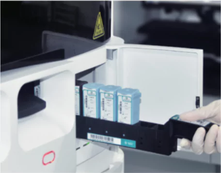
The Full Automatic Multiplex IHC Stainer by Celnovte, identified by plate number 1, is a significant advancement for pathology labs. This state-of-the-art equipment streamlines immunohistochemistry (IHC) staining procedures and is designed to meet diagnostic requirements while enhancing result quality. The plate number 1 model allows labs to run staining protocols simultaneously, accommodating a wide range of primary antibodies suitable for oncological, autoimmune, and infectious disease markers. By automating stages of the IHC procedure, this stainer device is intended to simplify tasks for pathologists, allowing them to focus on analyzing manual steps.
Features of CNT300 for Enhancing Accuracy
The CNT300 is equipped with enhancements that improve the accuracy of IHC outcomes. It includes a temperature regulation system that maintains consistent temperatures during staining to enhance antibody binding and reduce background staining levels. The automated reagent dispensation reduces the likelihood of errors and ensures accurate concentrations for consistent results. Additionally, the stainer incorporates quality control procedures to regularly validate machine performance, thereby maintaining result reliability and quality over time. This specific focus enhances precision and boosts trust in medical assessments.
Efficiency Benefits for Pathologists Using CNT300
In today’s fast-paced pathology labs, efficiency is of utmost importance. That’s why the CNT300 is the perfect solution. With its time-saving benefits, this automated immunohistochemistry staining machine can handle up to 60 slides at once and complete staining tasks in just 2.5 hours. By automating tasks like washing and applying reagents, it reduces labor and ensures consistency and accuracy in staining procedures. This is crucial in healthcare settings where quick access to vital diagnostic information is essential. Not only does it minimize waiting time for patients, but it also provides healthcare professionals with rapid access to crucial diagnostic data. It’s a win-win situation for everyone involved! By incorporating the CNT300 into your lab setup, you not only enhance the IHC staining procedure but also provide accurate and efficient services quickly and reliably. This ultimately improves the overall diagnostic process and enhances patient care.
What Are the Latest Advances in IHC Technology?
The advancements in automated staining technologies have been remarkable, with products like the Plate Number 1 Automatic Multiplex IHC Stainer by Celnovte transforming the realm of immunohistochemistry (IHC). Plate Number 1 provides adaptability by enabling the handling of multiple slides using diverse staining methods, which is crucial for labs managing large sample loads. This feature notably cuts down processing times and improves the consistency of staining outcomes. Additionally, the stainer incorporates software to oversee and regulate all aspects of the procedure, guaranteeing reliable outcomes that adhere to strict diagnostic criteria.
Innovations in Automated Stainers like the CNT300
A major advancement involves integrating temperature control systems into automated stainers, such as the CNT300 model. These systems ensure optimal conditions during the staining process, enhancing the effectiveness of antibody binding crucial for producing high-quality IHC stains. Automating handling and washing steps reduces errors and enables pathologists to achieve accurate results with minimal manual involvement. This automation boosts efficiency and introduces a quality control measure in clinical environments.
Future Trends in Multiplexing Techniques
As technology continues to advance, the use of techniques in IHC staining is becoming increasingly important. These methods allow pathologists to simultaneously visualize proteins in a tissue sample, providing a detailed look at the cellular environment. This feature is especially valuable in cancer care, as the interactions between biomarkers can impact treatment choices and prognosis assessments. It is expected that upcoming trends will focus on improving techniques further, potentially by introducing better imaging tools for clearer and more precise protein detection.
Pathologists are increasingly seeking tailored panels for diagnostic needs, integrating AI-powered tools for improved staining outcomes and automated measurements. Companies like Celnovte are advancing stainer technologies to enhance lab capabilities. These advancements in IHC technology have the potential to improve patient outcomes through precise and prompt diagnoses.
 Kit")

, MMab")
-IHC primary antibody")