 PRODUCT CATEGORY
CONTACT US


6 Cuizhu St, Zhong Yuan Qu, Zheng Zhou Shi, He Nan Sheng, China, 450001
Reticular Fiber Staining Kit
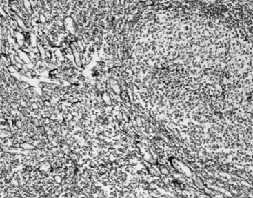
Reticular fibers are widely distributed and exist in two forms. One is a reticular scaffold for certain organs, such as bone marrow, spleen, lymph nodes, liver, thymus, and tonsils. The other is in the basement membrane of the epithelium. Meanwhile, smooth muscle, fat cells, capillaries, and nerve fibers are all covered with reticular fibers. There are no reticular fibers in the middle of the cancer tissue, but a large number of reticular fibros can be seen around its edge commonly known as cancer nests. Therefore, the reticular fibro staining is widely used in pathological diagnosis.
Product Features
- Staining Principle
Reticular fiber, a special type of collagen, are difficult to identify with ordinary HE staining and show argyrophilia. The silver ammonia solution is absorbed by the tissue and binds to the protein in the tissue, reduced to black metal silver by formaldehyde and deposited in the tissue and on the surface. After toning with gold chloride, the unreduced silver salt is washed away with sodium thiosulfate solution to make the reticular fibers in the tissue clearly showed. It can comprehensively display the damage details of reticular scaffold in the pathological tissue.
- Product Advantages
a)Clear organization structure, no obvious background staining
b)Stored for a long time
c)No precipitates and insoluble substances in each component reagent; Great stability
Specification
Components
|
Name |
Main Component |
|
|
Solution A: potassium permanganate solution |
Potassium permanganate |
|
|
Reagent (B) oxalic acid aqueous solution |
Oxalic acid |
|
|
Solution C: ammonium ferric sulfate solution |
Ammonium ferric sulfate |
|
|
Solution D: tollens solution |
Silver nitrate, ammonia water |
|
|
Solution E: formaldehyde solution |
Formaldehyde |
|
|
Solution F: gold chloride solution |
Gold chloride |
|
|
Solution G: sodium thiosulfate solution |
Sodium thiosulfate |
|
More Info
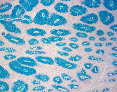
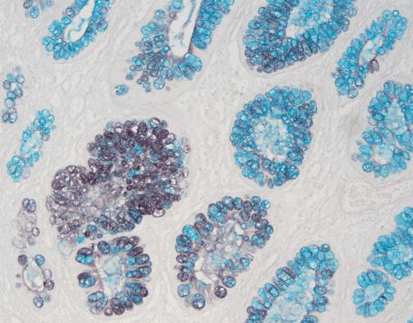
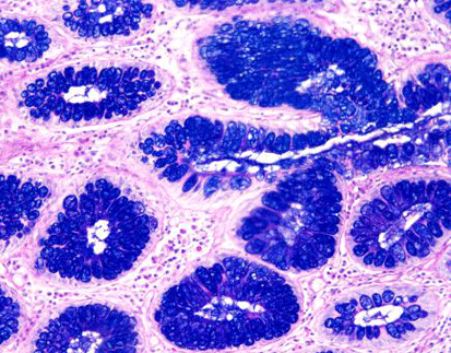
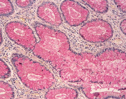
- Staining Results : acidic mucus material is blue, neutral mucus material is red, mixed mucus material is blue-violet, and cell nuclei are light blue
- Specifications: 7×10mL/kit, 7×20mL/kit,7×100mL/kit
- Storage: 2-8 ℃
- Shelf Life: 18 months
Related Product


 PRODUCT CATEGORY


 Chat
Chat

 message

 Quote
Quote
