
-
Home
-
Product

-
IMMUNOHISTOCHEMISTRY
-
INSTRUMENT
-
MOLECULAR
-
HISTOLOGY
-
CYTOLOGY
-
DIGITAL PATHOLOGY
-
-
Solution
-
Support
-
About
-
Contact

Top 10 Special Stains Every Dermatologist Should Know
Are you eager to learn about special stainingin dermatology? Jump into this blog to explore mucin, amyloid, and beyond—unveiling their diagnostic strength! Discover expenses, perks, and advanced solutions. Sharpen your skin analysis skills—read now!
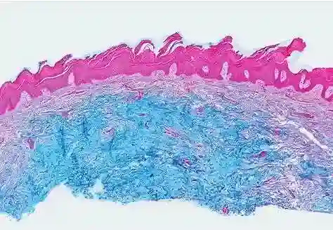
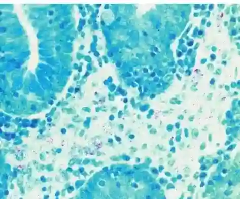
1. Mucous staining
Mucous staining is a vital method in dermatology. It lets you see mucins in tissue samples clearly. Mucins are glycoproteins that play a key part in many bodily and disease processes. These include lubrication and shielding of epithelial surfaces. In dermatology, mucous staining helps spot conditions marked by too much or odd mucin buildup.
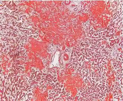
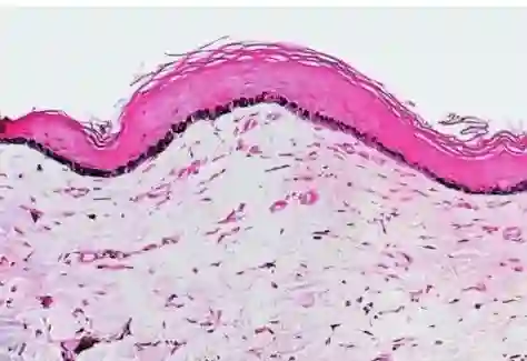
2. Amyloid substance
Amyloid substance staining is essential for finding amyloid deposits in tissues. These link to a variety of widespread and local ailments. The deposits are made of misfolded proteins that clump into tough fibers. Spotting amyloid deposits aids in diagnosing issues like primary skin amyloidosis and widespread amyloidosis.
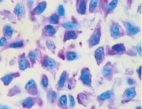
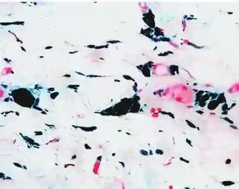
3. Mast cell staining
Mast cells are crucial to immune reactions. They have a big role in allergic responses and other skin conditions. Mast cell staining lets dermatologists view these cells in tissue samples plainly. It offers clues about disorders like urticaria pigmentosa and mastocytosis. This method also helps gauge mast cell numbers and spread.
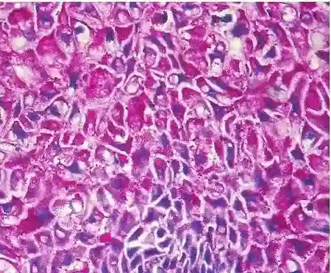
4. Glycogen staining
Glycogen is a sugar compound that stores energy in cells. Glycogen staining is used to detect its deposits in tissue samples. These can hint at metabolic issues or other disease states. The technique proves especially handy in diagnosing glycogen storage problems affecting the skin.
5. Acid-fast staining
Acid-fast staining is applied to spot acid-fast bacilli, like Mycobacterium types, in tissue samples. This approach is critical for diagnosing infectious diseases such as skin tuberculosis and leprosy. These show distinct skin signs. Accurately finding these germs supports timely diagnosis and care.
6. Melanin staining
Melanin is the pigment that gives skin its hue. Its spread can shift in various skin disorders. Melanin staining allows clear viewing of melanin in tissues. This helps diagnose pigment issues like vitiligo, melasma, and melanoma.
7. Iron staining
Iron buildup in tissues can stem from various disease processes, like hemochromatosis and hemosiderosis. Iron staining methods reveal hemosiderin deposits in skin biopsies. This provides useful info for diagnosing conditions tied to odd iron handling.
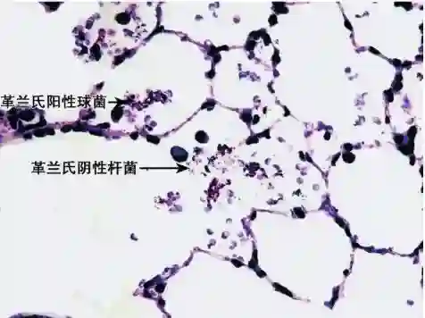
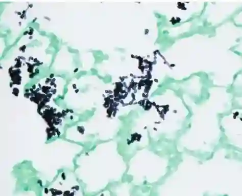
8. Bacterial staining
Bacterial infections can show up with varied skin signs that need precise spotting for proper treatment. Bacterial staining methods let you see germs in tissue samples distinctly. This eases diagnosis of infections from bacteria like Staphylococcus aureus or Streptococcus pyogenes.
Incorporating special stains into dermatology work boosts diagnostic precision. It gives deep insights into the makeup of skin tissues at cellular and molecular levels.
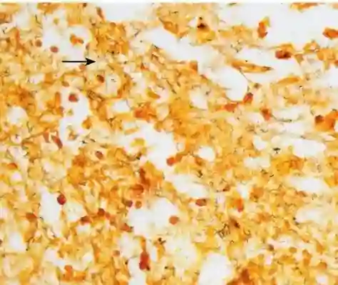
9. Fungal staining
Fungal infections are common in dermatology. They demand exact identification for effective treatment. Fungal staining methods aim to highlight fungal parts in tissue samples vividly. This enables dermatologists to diagnose conditions like dermatophytosis and candidiasis. These stains sharpen the view of fungi. They also aid in telling apart different fungal types.
10. Spirochaeta staining
Spirochaetes are a group of germs that can trigger major skin conditions, including syphilis and Lyme disease. Spirochaeta staining is key for spotting these spiral-shaped germs in tissue samples. This method ensures accurate detection and diagnosis. It supports prompt action and handling of spirochaetal infections.
Importance of Special Staining in Dermatology
Special staining methods play a central role in dermatology. They offer sharper diagnostic skills.
Enhancing Diagnostic Accuracy
Special stains provide thorough insights into skin tissues’ cellular and molecular makeup. This improves diagnostic precision greatly. They let you see specific cell parts or germs that routine stains might miss. Such clarity helps in correctly diagnosing and managing various skin issues.
Common Applications in Dermatology
In dermatology, special stains are often used to pinpoint infectious agents. They also detect odd deposits and show specific cell structures clearly. These uses allow dermatologists to diagnose a broad array of skin problems accurately and swiftly.
Choosing the Right Stain for Accurate Diagnosis
Picking the proper stain is crucial for getting spot-on diagnostic results.
Factors Influencing Stain Selection
Several factors sway stain choice. These include the suspected issue, tissue kind, and the target you want to see. Grasping these elements helps dermatologists select the best stain for each case. This ensures top diagnostic outcomes.
Integrating Multiple Stains for Comprehensive Analysis
Using several stains together can offer a full analysis. It highlights different sample aspects at once. Methods like multi-color immunohistochemistry let you watch multiple targets on one slide simultaneously. This gives insights into complex tissue interactions.
Celnovte: A Reliable Dermatology Staining Solution Supplier
Celnovte is devoted to delivering top-notch staining solutions. These meet dermatologists’ needs worldwide.
Overview of Celnovte’s Product Range
Celnovte offers a wide lineup of products. These are crafted to back various staining methods in dermatology. Their range includes fully automated staining setups. These ensure steady and trustworthy results.
Commitment to Quality and Innovation
Celnovte puts quality and fresh ideas first. They keep crafting new tools to boost staining methods. Their devotion ensures dermatologists get cutting-edge solutions. These sharpen diagnostic precision and patient results.
FAQs on Special Staining in Dermatology
What are the benefits of using special stains over routine stains?
Special stains offer clearer views of specific cell parts or germs. This leads to more exact diagnoses than routine stains provide. They allow targeted spotting of oddities that standard methods might not show.
How do I determine which stain to use for a particular condition?
Figuring out the right stain means weighing factors like the suspected issue, tissue type, and target to view. Chatting with skilled pathologists helps. So does tapping resources from trusted suppliers like Senot Biotech. They offer a full immunohistochemistry solution with over 460 primary antibodies. This aids in picking the best stain.
Can special staining techniques be used on all types of skin samples?
Special staining methods can work on many skin samples. Yet, factors like tissue keeping and prep can sway how well they do. Following top practices for handling tissues is key. This ensures the best results from special stains.
Senot Biotech delivers a full immunohistochemistry solution. They provide top-quality results. These boost the power of special stains used by dermatologists worldwide.

