The Meaning of the IHC Markers
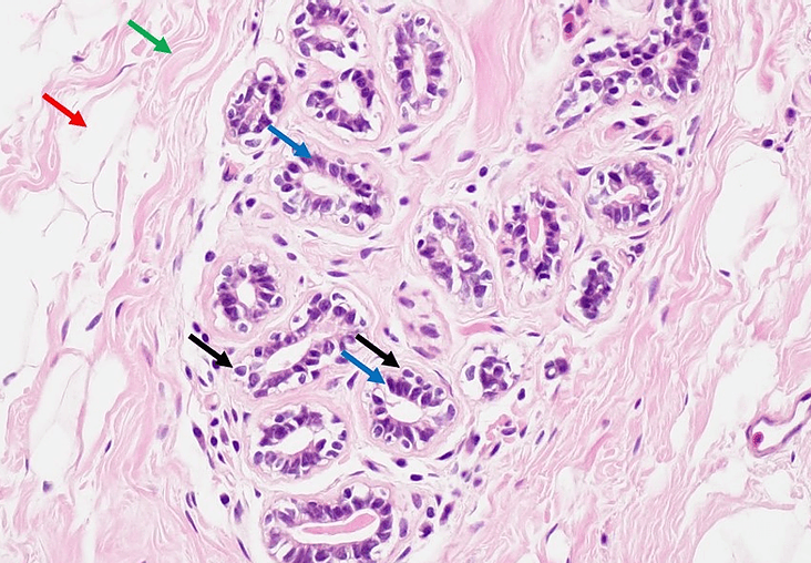
Briefly, I would like to give a couple examples of typical IHC stains and what they mean. For example, below is a hematoxylin and eosin (H&E) image of normal breast tissue. For the most part, breast tissue is composed of ductal/lobular epithelium (blue arrows) surrounded by mostly fat (adipose) tissue (red arrow) and collagen fibers (green arrow). Collagen is produced by fibroblasts, cells with flattened nuclei seen distributed among the collagen. Myoepithelial cells (black arrows) are cells located beneath the simple columnar ductal epithelium, but within the basement membrane of the ducts. Myoepithelial cells are contractile cells and form a discontinuous layer surrounding the epithelium. Their function is to constrict the ducts during lactation to expel milk. They appear similar to the glandular cells, but in this image, most of these cells have a clear ring around the nuclei.
Breast cancer is a very common type of tumor and treatment depends greatly on how the tumor cells behave. To that end, typical IHC stains performed are estrogen receptor (ER), progesterone receptor (PR), and human epithelial growth factor receptor 2 (HER2).
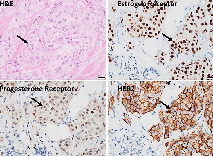
All normal female-related tissues such as breast, uterus, and ovaries express ER and PR. These receptors are found on the nucleus and cause the cell to respond in the presence of estrogen and progesterone. ER and PR are nuclear stains. In the IHC serial sections below, brown color is positive staining and blue is negative staining. These images represent normal control tissues. As can be seen, not every nucleus stains positively, but many do which is the typical positive staining pattern. The 4th image below shows HER2 1+ staining on normal breast tissue. HER2 is a membrane receptor and is constitutively expressed at low levels in many body tissues. The best HER2 controls are composed of at least 4 tissues expressing HER2 at various levels (0, 1+, 2+, 3+).
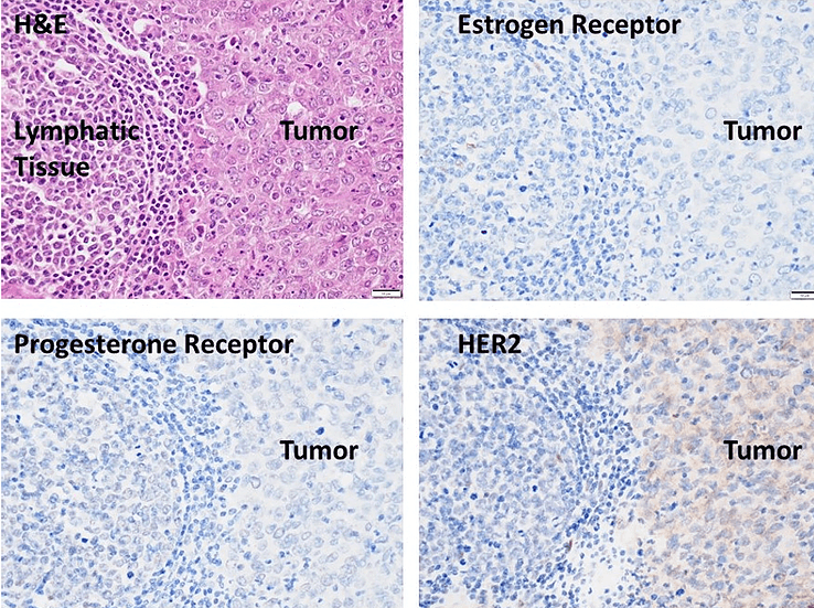
Below is an example of a ductal breast adenocarcinoma. In each of the serial sections, there is benign lymphatic tissue on the left and dense tumor on the right. As can be seen, there is no staining of the tumor cells for ER and PR. What that indicates is that using hormonal suppression as a cancer therapy would not be effective in this patient. HER2 is expressed in low levels corresponding to 1+. Grades of 0 and 1+ are considered negative for overexpression. A 2+ is equivocal, and a 3+ is positive for overexpression. Although HER2 3+ tumors are typically more aggressive, there is a treatment, Trastuzumab (Herceptin), which targets HER2 specifically and is very effective against the 3+ tumors.
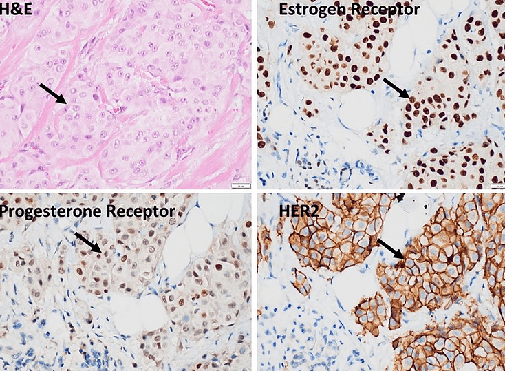
In the set of images below, the tumor is shown by the black arrow. As can be seen, many nuclei in the ER and PR stained sections are positive which indicates that this tumor is very responsive to estrogen and progesterone so suppressing those hormones would most likely suppress tumor growth. HER2 is staining 3+ so this tumor would also be responsive to Trastuzumab.
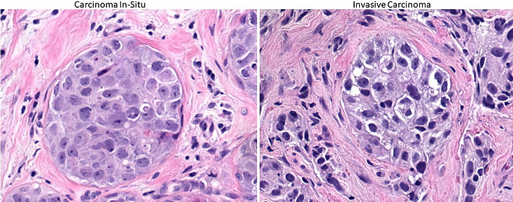
Most of the time, a breast cancer can be classified as in-situ (not broken through the basement membrane, yet) or invasive by examining H&E alone. See images below. In-situ groups of tumor cells, like benign epithelium, have surrounding myoepithelial cells and tend to have smooth borders with a well-defined ring of collagen surrounding the duct. See left image below. Invasive tumor is typically less well circumscribed and cells can be seen burrowing into neighboring collagen.
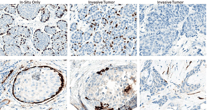
When the distinction between in-situ and invasive tumor is unclear, two markers can be used to make the diagnosis. P63 is a myoepithelial cell marker and smooth muscle myosin highlights the basement membrane. When an “unbroken” ring of myoepithelial cells or basement membrane surrounds the tumor cells such as is seen in the two left images below, the tumor is in-situ. However, if there is discontinuity of the myoepithelial ring or the basement membrane, the tumor is invasive (See 4 right images below). Treatment for invasive tumor is, of course, more aggressive.
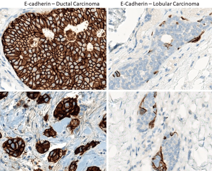
Most breast cancers stem from the ductal epithelium or epithelium of the milk-producing lobes themselves. Typically, this distinction is seen on H&E morphology. However, there are some instances where the tumor type is not clear and the pathologist wants to verify the diagnosis. E-cadherin is a marker which distinguishes between the two. E-cadherin is uniformly positive in ductal carcinomas (left images below) and negative in lobular carcinomas (right images below). Although some positive staining can be seen in the lobular carcinomas, the cells staining positively represent ductal cells which are still attached to the malignant lobular epithelium.
From Dr. Chen Shichang (Pathology technology exchange)
Related News


 2021-02-24