 Home
Home
 Product
 IMMUNOHISTOCHEMISTRY
 MULTIPLEX IHC
 Calponin/p63 detection reagent (Immunohistochemical)
  PRODUCT CATEGORY
CONTACT US


6 Cuizhu St, Zhong Yuan Qu, Zheng Zhou Shi, He Nan Sheng, China, 450001
Calponin/p63 detection reagent (Immunohistochemical)
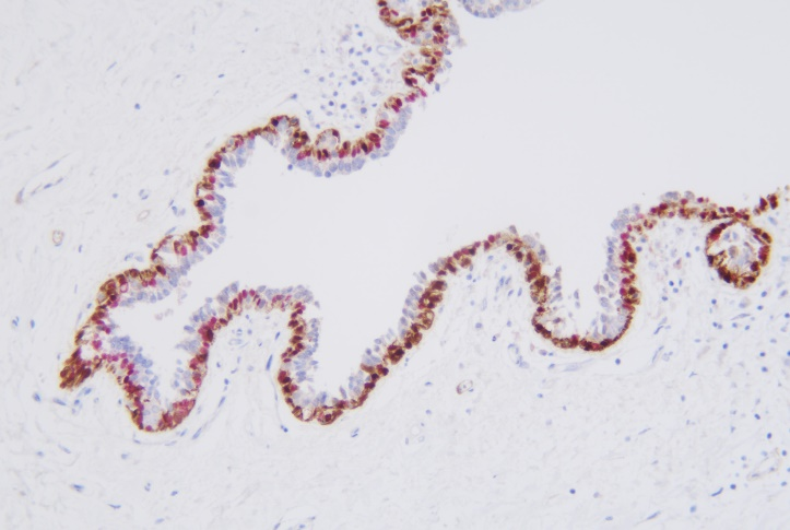
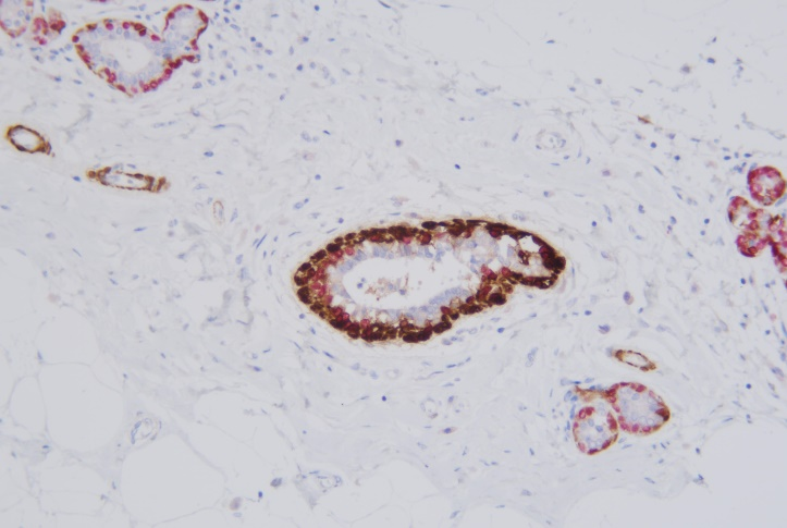
Calponin/p63 detection reagent (Immunohistochemical) is mainly used to qualitatively identify Calponin and p63 proteins. Using immunohistochemical dual staining detection and multiple antibody combination technology, there are detected two antigen targets at the same time on the same sample tissue section by one staining process.
Specification
|
Model Number |
Specification |
|
SD8106 |
1mL/pcs; 3mL/pcs; 7/pcs |
This antibody is used with Immune Chromogenic Reagent (Dual Staining I) (SD8001)
Clone:C6A12 + BP6038
More Info
Calponin is expressed in differentiated and mature smooth muscle cells, and shows strongly positive in vascular smooth muscle cells. Calponin shows strongly positive in myoepithelial cells in UDH, ADH, and DCIS. Compared with SMA, it has strong specificity and less cross-reactivity to myofibroblasts than SMA and MSA.
p63 is a member of p53 gene family and a better marker for breast myoepithelial cells. It has strong specificity and sensitivity and can accurately display myoepithelial cell nucleus. Because p63 shows negative in myofibroblasts, it is more superior than SMA and Calponin.
When immunohistochemical staining does not show myoepithelium around the tumor, it supports the diagnosis of interstitial infiltration. However, it is difficult to determine whether myoepithelial cells are truly lacking or false (caused by severe squeezing or cutting problems). The following features support the true lack of myoepithelial cells: ① Myoepithelial cells are not detected around the middle-large or multiple tumor cell nests; ② There is no expression of two different myoepithelial cell markers. Therefore, Calponin and p63 are good complementary markers.
Breast Normal Duct-p63-nucleus-red
Breast Normal Duct Calponin–cytoplasm-brown
Related Product


 PRODUCT CATEGORY


 Chat
Chat

 message

 Quote
Quote


