 PRODUCT CATEGORY
CONTACT US
Product Features
Application
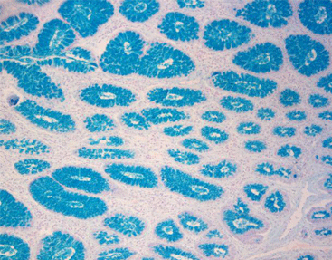
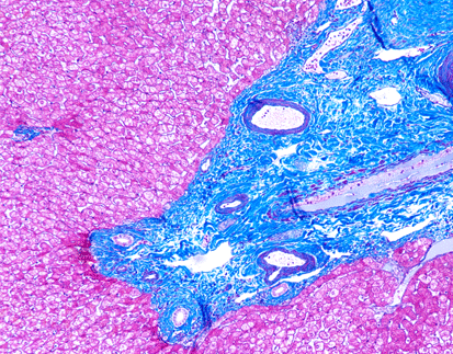
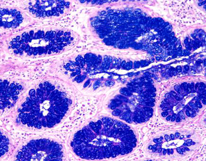
The Alcian Blue (pH 2.5) Stain Kit is intended for use in the histological visualization of sulfated and carboxylated acid mucopolysaccharides and sulfated and carboxylated sialomucins (glycoproteins).
Staining Principle
Alcian blue is the most specific stain showing acidic mucus substances. This cationic dye forms salt linkage with acidic groups. The different pH values of the stain liquid can be used to distinguish the mucous substance. When the pH is 1, the carboxyl group (-COOH) is not stained, and the sulfonic acid group (-SO3H) is stained. When the pH is 2.5, the carboxyl group stained well but the sulfuric acid mucus stained.
Product Advantages
a) Bright staining Color
b) No precipitates and insoluble substances in each component reagent; Great stability.
Suggested Controls (not provided)
– Small Intestine, Appendix, Colon
Procedure
1. Deparaffinize sections if necessary and hydrate to distilled water.
2. Incubate slide in Acetic Acid solution for 3 minutes.
3. Stain tissue section with Alcian Blue Solution (pH 2.5) solution for 30 minutes at room temperature or 15 minutes at 37° C.
4. If desired, rinse slide briefly in Acetic Acid solution to remove excess Alcian Blue.
5. Rinse for 2 minutes in running tap water followed by 2 changes of distilled water.
6. Stain tissue section with Nuclear Fast Red Solution (Enhanced Stability) for 5 minutes.
7. Rinse for 2 minutes in running tap water followed by 2 changes of distilled water.
8. Dehydrate through graded alcohols.
9. Clear, and mount in synthetic resin.
References
1. Hoshino‐Negishi K, Ohkuro M, Nakatani T, Kuboi Y, Nishimura M, Ida Y, Kakuta J, Hamaguchi A, Kumai M, Kamisako T, Sugiyama F. Role of Anti‐Fractalkine Antibody in Suppression of Joint Destruction by Inhibiting Migration of Osteoclast Precursors to the Synovium in Experimental Arthritis. Arthritis & Rheumatology. 2019 Feb;71(2):222- 31.
2. Li B, Lee C, Martin Z, Li X, Koike Y, Hock A, Zani-Ruttenstock E, Zani A, Pierro A. Intestinal epithelial injury induced by maternal separation is protected by hydrogen sulfide. Journal of Pediatric Surgery. 2016 Oct 26.
3. Li B, et al, Endoplasmic reticulum stress is involved in the colonic epithelium damage induced by maternal separation, J Pediatr Surg (2016)
4. Kumar G, Hara H, Long C, Shaikh H, Ayares D, Cooper DK, Ezzelarab M. Adipose-derived mesenchymal stromal cells from genetically modified pigs: immunogenicity and immune modulatory properties. Cytotherapy. 2012 Apr 1;14(4):494-504.
5. Lillie, R.D. 1977, H.J. Conn’s Biological Stains, 9th Edition. Williams & Wilkins, Baltimore. Pages 452-455.
6. Sheenan, D.C., Hrapchak, B.B. Theory and Practice of Histotechnology, 2nd Edition. Battelle Press, Columbus, OH. Pages 172-173.
7. Churukian, C.J., 1989, Manual of Special Stains Laboratory, 4th Edition. University of Rochester, Rochester, New York. Pages 55-56.
8. Carson, F.L., 1996, Histotechnology; A Self-Instructional Text, 2nd Edition. ASCP Press, Chicago, IL. Pages 117-121.
9. Leow, C.C., Romero, M.S., Ross, S., Polakis, P., and Gao, WQ. Hath1, Down-Regulated in Colon Adenocarcinomas, Inhibits Proliferation and Tumorigenesis of Colon Cancer Cells. Cancer Research 64, 6050-6057, September 1, 2004.
Specification
|
Main Component |
|
Alcian blue, glacial acetic acid |
|
Nuclear fast red |
More Info
- Storage:2-8 ℃
- Shelf Life: 18 months
Related Product


 Chat
Chat

 message

 Quote
Quote


